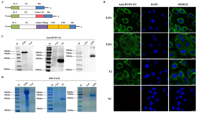
Figure 1.
Immuno-expression and identification of E2, E2Ft, and E2Fc proteins in HEK293 cells. (A) The construction diagrams of BVDV-E2, BVDV-E2Ft, and BVDV-E2Fc. (B) Detection of the immuno-expression of recombinant proteins by IFA; 48 h after transfected with E2, E2Fc, or E2Ft, HEK293 cells were fixed for IFA. BVDV E2 monoclonal antibody and FITC-goat anti-mouse IgG were applied as primary and secondary antibody, respectively. Nucleus was stained with DAPI (blue). Scale bar represents 20 μm. (C) Detection of purified recombinant proteins by Western blot. (D) Identification of purified recombinant proteins by SDS-PAGE.

