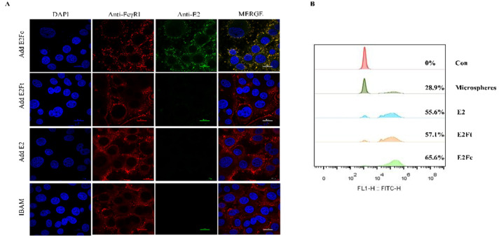
Figure 2.
Activation of the APC-FcγRI by BVDV-E2Fc. (A) Co-localization analysis of CD64 (FcγRI) with E2Fc protein on BAM was detected by confocal microscopy. CD64 (FcγRI) on BAM was stained with rabbit anti-CD64 and Cy3-labeled goat anti-rabbit IgG as the primary and secondary antibody, respectively. E2Fc was stained with mouse anti-E2 monoclonal antibody and Alexa Fluor® 488 goat anti-mouse IgG as the primary and secondary antibody, respectively. Nucleus was stained with DAPI (blue). Images were merged to analysis the co-localization (yellow) of CD64 (FcγRI) with E2Fc. Scale bar represents 20 μm. (B) Flow cytometry analysis of phagocytosis rate of E2 proteins on BAM. BAM were pre-incubated with E2, E2Ft, or E2Fc protein for 4 h. Cells were incubated with 10 μL FITC-conjugated microspheres for 2 h. Adherent macrophages were digested with trypsin and resuspended with PBS. Fluorescence intensity at 488 nm was determined by the Beckman flow cytometer.

