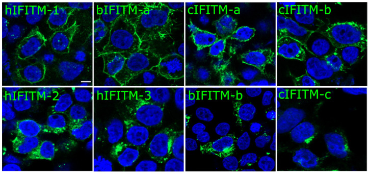
Figure 3.
Cellular localization of human, bovine, and camel IFITMs, as indicated. HEK293T cells were transfected with plasmids encoding the different IFITMs. Twenty-four hours post-transfection, the cells were fixed with PFA and immuno-stained for IFITMs with an anti-HA antibody followed by a secondary antibody conjugated to Alexa488 (Green). Cell nuclei were labelled with DAPI staining (blue). Representative pictures of IFITMs expressing cells are shown. The scale bar in the upper left panel represents 5 µm. hIFITM-1, human IFITM-1; hIFITM-2, human IFITM-2; hIFITM-3, human IFITM-3; bIFITM-a, bovine IFITM-a; bIFITM-b, bovine IFITM-b; cIFITM-a, camel IFITM-a; cIFITM-b, camel IFITM-b; cIFITM-c, camel IFITM-c.

