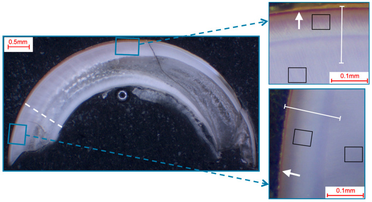
Figure 1.
Selection of fields and line-scans for the elemental analysis in murine incisors shown in a photo-microscopic overview. The black squares indicate the 49 µm × 39 µm areas of the field scans, while the white lines indicate the paths of the line scans (length: approximately 140 µm). The white dotted line shows the pre/post-eruptive border, with the pre-eruptive scan area being enlarged in the top right and the post-eruptive scan area being enlarged in the bottom right picture. All pictures showed a yellow-brownish enamel, which is indicative of iron in the superficial enamel (white arrows).

