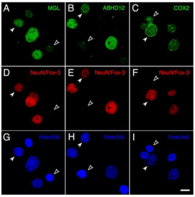
Figure 6.
Panoramic micrographs of intact nuclei isolated from the adult rat brain cortex processed for MGL-, ABHD12- and COX2-immunofluorescence (A–C) combined with NeuN/Fox-3-immunofluorescence (D–F) and Hoechst’s chromatin staining (G–I). Every NeuN-positive nucleus exhibited strong immunoreactivity for MGL, ABHD12 and COX2 (filled arrowheads), whereas NeuN-negative nuclei (empty arrowheads) displayed clear positive staining only for COX2. All micrographs were acquired in grayscale and pseudo colored. Scale bar = 20 µm in I (applies to (A–I)).

