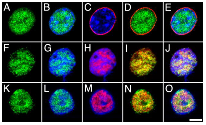
Figure 7.
High-power micrographs of intact nuclei isolated from the adult rat brain cortex double stained with anti-MGL (green) combined with Hoechst staining (blue) and NPCx ((A–E); red), NeuN/Fox-3 ((F–J); red) and DGLα ((K–O); red) immunostaining. All micrographs are maximum intensity projections of three consecutive 0.24 µm-thick optical sections acquired in grayscale and pseudo colored. Scale bar = 5 µm in O (applies to (A–O)).

