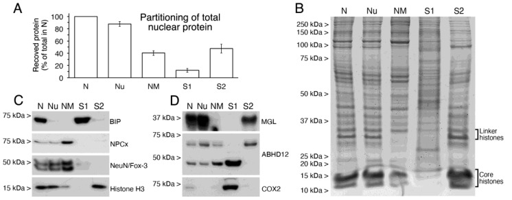
Figure 10.
Characterization of nuclear subfractions obtained from intact nuclei and analysis of nuclear compartmentation of MGL, ABHD12, COX2. (A) Bar graph depicting the partitioning of total protein from cortical intact nuclei (N) into nucleoids (Nu), nuclear matrix (NM), TX-100 extractable supernatant (S1) and DNase I/high salt extractable supernatant (S2). Data are expressed as percent protein recovered in each fraction relative to total protein (100%) in N fraction. Values are mean ± SD of two independent experiments. (B) Coomassie blue-stained SDS-PAGE of equivalent amounts (20 µg) of protein from N, Nu, NM, S1 and S2 samples. (C) Western blot analysis of nuclear subfractions (12 µg/lane) run in parallel to analyze the suitability of the subfractioning procedure using specific markers for non-ionic detergent extractable (BiP), non-ionic detergent and DNase I/high salt resistant (NPCx), nuclear matrix (NeuN/Fox-3) and DNA-bound (Histone H3) proteins. (D) MGL, ABHD12 and COX2 immunoblots in N, Nu, NM, S1 and S2 nuclear subfractions. 12 µg/lane were loaded.

