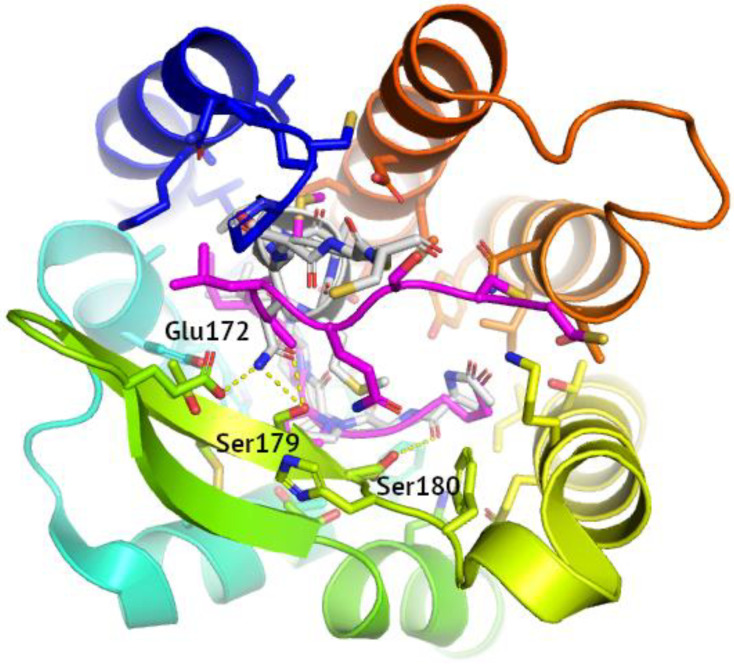
Figure 2.
Reconstruction of ligand binding mode in 5UIW self-docking. The reference PDB pose of the 5UIW ligand was shown in grey, with the polar contacts involving side chains indicated with yellow dashed lines. The residues involved in polar contacts located in ECL2 were labeled. The Glide-reconstructed ligand pose was shown in magenta (RMSD equal to 4.19 Å).

