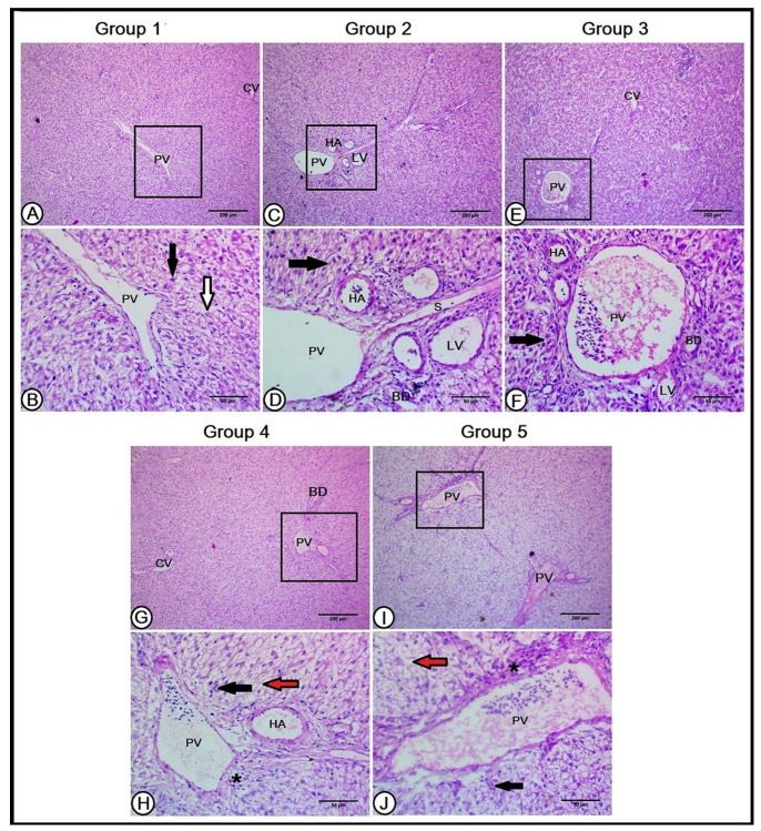
Figure 1.
A histopathological analysis of the hepatic architecture in the experimental groups (A,B): the livers in the first group (Con) showed normal hepatic architecture. The central vein (CV) was in the center of each hepatic lobule and the hepatocytes (black arrow) were arranged in plates that radiate longitudinally outward from the central vein that was separated by a hepatic sinusoid (white arrow). The portal triad contained a branch of the portal vein (PV). The livers of the second and third groups (C–F, respectively) revealed a better organization of the hepatic cells (black arrows) and hepatic sinusoid (S) in between. The portal triad is composed of a large-sized branch of the portal vein (PV), with numerous branches of the hepatic artery (HA), and bile ducts (BD), in addition to lymphatic tissues (LV). The livers in the fourth and fifth groups (G–J, respectively) illustrate disorganized and degenerated hepatocytes (black arrows) around the central vein (CV), numerous variable-sized hepatic sinusoids (red arrows), excessive connective tissue, lymphocytic infiltration (black asterisks), fewer branches of the hepatic artery (HA), and bile duct (BD).

