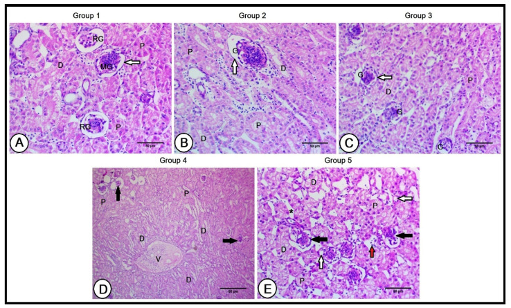
Figure 2.
A histopathological analysis of the renal cortex in the experimental groups (A): Renal cortex of the control group showing glomeruli of reptilian-type nephrons (RG) and glomeruli of mammalian nephrons (MG), and proximal (P) and distal (D) convoluted tubules. The glomerulus was surrounded by thin glomerular basement membranes (black arrow). The renal cortex of the second (B) and third (C) groups revealed large-sized proximal (P) and distal (D) convoluted tubules. The glomeruli (G) increased in size in the second group; however, their size was slightly reduced in the third group. The glomeruli were surrounded by thin glomerular basement membranes. The renal cortex of the fourth group (D) shows small-sized glomeruli (black arrows) and a concentric arrangement of distal convoluted tubules (D) around the intralobular vein (V). The renal cortex of the fifth group (E), illustrating small-sized glomeruli, surrounded by irregularly disrupted basement membranes (black arrows), and large interstitial spaces (black asterisk) between small-sized proximal (P) and distal (D) convoluted tubules. Note the presence of atrophied tubules (white arrows) and few lymphocytic infiltrations (red arrows).

