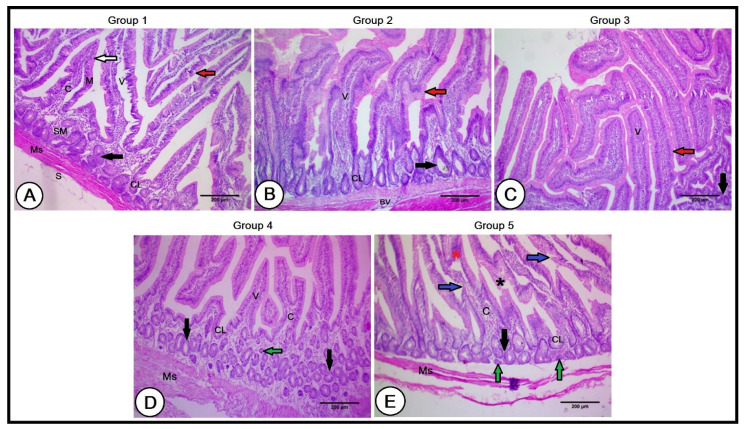
Figure 3.
A histopathological analysis of the small intestines in the experimental groups (A): Duodenum of the control group showing mucosa (M), submucosa (SM), muscularis (Ms), and serosa (S) tunics. The lamina epithelialis consisted of simple columnar epithelial cells (white arrows) and goblet cells scattered among them (red arrows). The intestinal villi consisted of connective tissue core (C) and exhibited intestinal glands (crypts of Lieberkühn, CL). The submucosa revealed submucosal glands (black arrows). (B,C): Intestinal wall of the second and third groups, respectively, revealing longer and wider intestinal villi (V), numerous goblet cells (red arrows), deeper and larger crypts of Lieberkühn (CL), and large-sized submucosal glands in the second group, with the beginning of their size reduction in the third group (black arrows). (D): Duodenal wall of the fourth group showing reduced intestinal villi (V) packed with a less connective tissue core (C). The intestinal glands (CL) were smaller and the submucosa glands were numerous and small in their size (black arrows). (E): Duodenal wall of the fifth group illustrating short intestinal villi with irregular lamina epithelialis (blue arrows), degenerated areas (black asterisk), lymphocytic infiltration (red asterisk), poor connective tissue cores (C), and smallest and shortest intestinal glands (CL). Note the presence of numerous small-sized submucosal glands (black arrows), together with some atrophied ones (green arrows), and a narrow muscular tunic (Ms).

