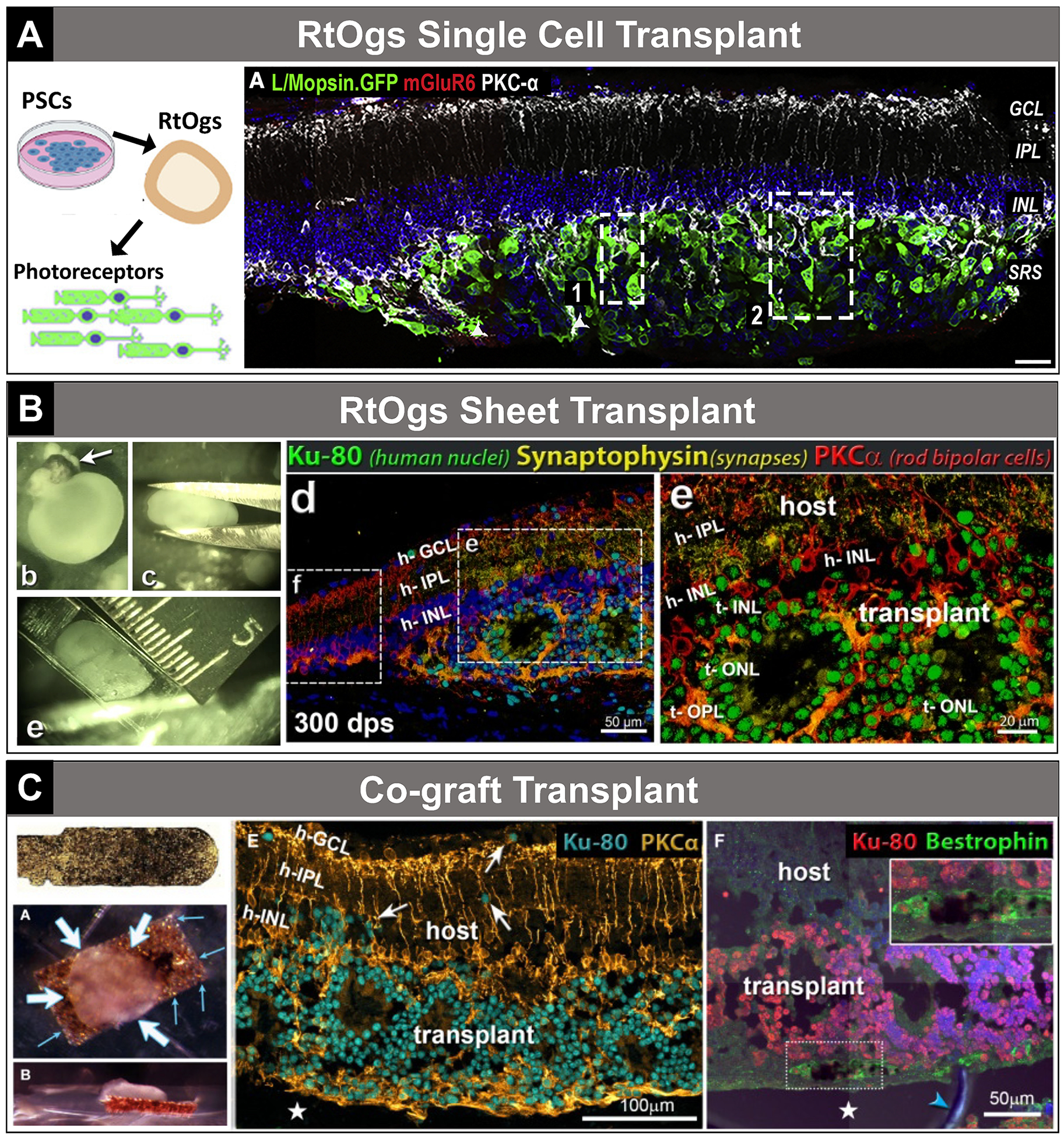
FIGURE 3.

Transplantation examples—single cell, sheet, cograft. A, Single-cell transplantation. Taken from Ribeiro et al39 (graphical abstract; Fig. 3A). B, Sheet transplantation. Taken from McLelland et al16 (Supplemental Fig. 1; Figs. D–E; republished with permission of Investigative Ophthalmology & Visual Sciences, from McLelland et al, Transplanted hESC-derived retina organoid sheets differentiate, integrate, and improve visual function in retinal degenerate rats. Invest Ophthalmol Vis Sci. 2018;59:2586–2603; doi:10.1167/iovs.17-23646; permission conveyed through Copyright Clearance Center, Inc). C, Cograft transplantation. Taken from Thomas et al45 (Fig. 1I; Figs. 3A–B; Figs. 7E–F). h-GCL indicates host ganglion cell layer; h-IPL, host inner plexiform layer; h-INL, host inner nuclear layer; PSCs, pluripotent stem cells; RtOgs, retinal organoids; t-INL, transplant inner nuclear layer; t-OPL, transplant outer plexiform layer; t-ONL, transplant outer nuclear layer.
