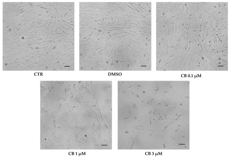
Figure 8.
Morphology of human Wharton’s jelly mesenchymal stem cells (hWJ-MSCs) after 24 h in the presence of Cytochalasin B (CB) 0.1 μM, 1 μM, and 3 μM, dimethyl sulfoxide (DMSO) 0.05% (CB vehicle) or without treatment (CTR). Images were acquired using the Leica Labovert FS Inverted Microscope equipped with a Leica MC170 HD Imaging System Camera. Scale bars: 50 μm.

