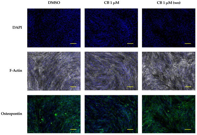
Figure 13.
Immunofluorescence analysis in human Wharton’s jelly mesenchymal stem cells (hWJ-MSCs) of cytoskeletal and osteogenic markers at 7 days from the beginning of the osteogenic program. hWJ-MSCs were cultured in osteogenic medium with dimethyl sulfoxide (DMSO) 0.05% (CB vehicle) or CB 1 μM for the entire experimental time (continuous CB). hWJ-MSCs were also cultured in osteogenic medium (OM) in presence of CB 1 μM for only 24 h, and then removed, and cells were cultured in OM up to the end of the experiment (wo = CB wash-out). hWJ-MSCs were immunostained with Phalloidin (grey signal, specific for F-Actin) or anti-osteopontin (green signal) at 7 days from the beginning of the osteogenic program. NucBlue® Fixed Cell ReadyProbes® Reagent (DAPI) was used to counter-stain nuclei (blue signal). Images were acquired using a Nikon inverted microscope Eclipse Ti2-E and a digital sight camera DS-Qi2, through the imaging software NIS-Elements. Scale bars: 100 μm.

