Figure 2.

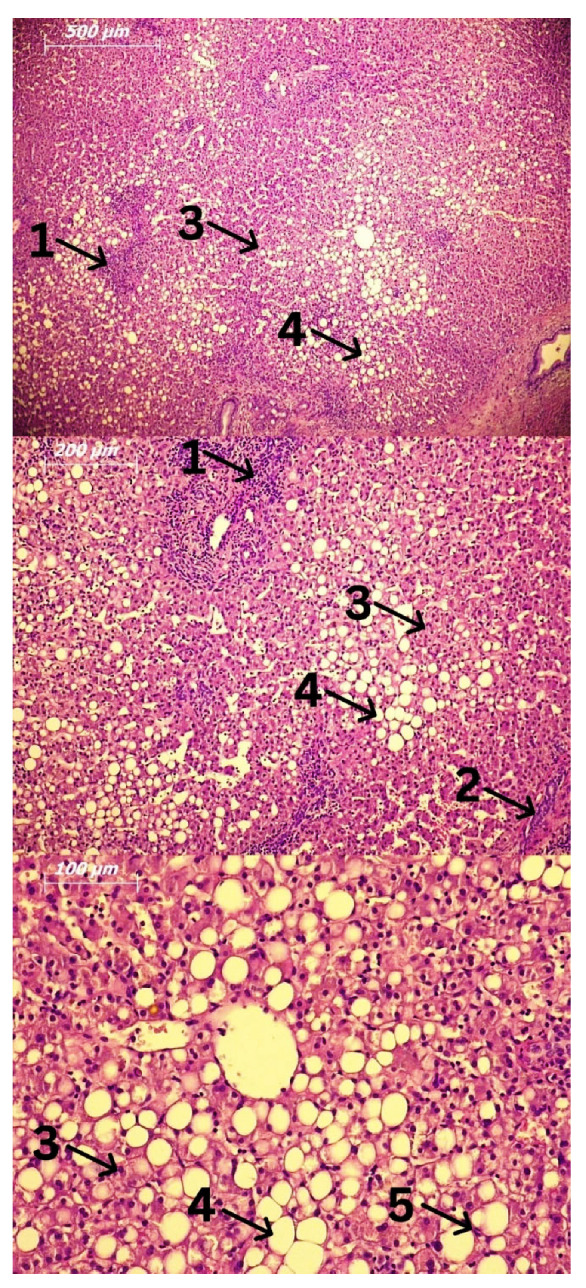
Fatty liver biopsy. Hematoxylin and Eosin (H&E) staining. Legend: 1: Lymphocytes; 2: Focal necrosis; 3: Ballooned hepatocytes; 4: Fat accumulation; 5: Kupffer cells.

Fatty liver biopsy. Hematoxylin and Eosin (H&E) staining. Legend: 1: Lymphocytes; 2: Focal necrosis; 3: Ballooned hepatocytes; 4: Fat accumulation; 5: Kupffer cells.