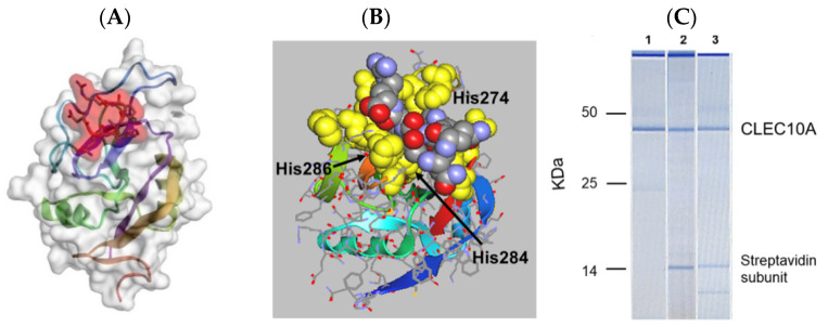
Figure 5.
(A) In silico docking of an arm of sv6D (NQHTPR) to human CLEC10A (accession no. 6PY1, RMSD = 1.343 Å, ΔG′ = −37 kJ/mol) with CABS-Dock. The peptide is enclosed in red shading. (B) The structure in (A) was downloaded into ArgusLab. The position of sv6D in the binding pocket is shown after additional molecular dynamics. The peptide is colored (carbon, grey; nitrogen, blue; oxygen, red) while the binding site is yellow. The positions of His274, His284, and His286 of the binding site are indicated. (C) A lysate of human monocyte-derived DCs was incubated with (1) mouse anti-human CLEC10A, which was recovered with magnetic beads coated with protein A; (2) biotinylated sv6D; or (3) biotinylated svL4, which were recovered with magnetic beads coated with streptavidin. Proteins were eluted from the beads and subjected to electrophoresis. Molecular mass markers are indicated for IgG heavy chain (50 kDa), IgG light chain (25 kDa), and a streptavidin C1 subunit (13.6 kDa). The top band is an instrument marker.

