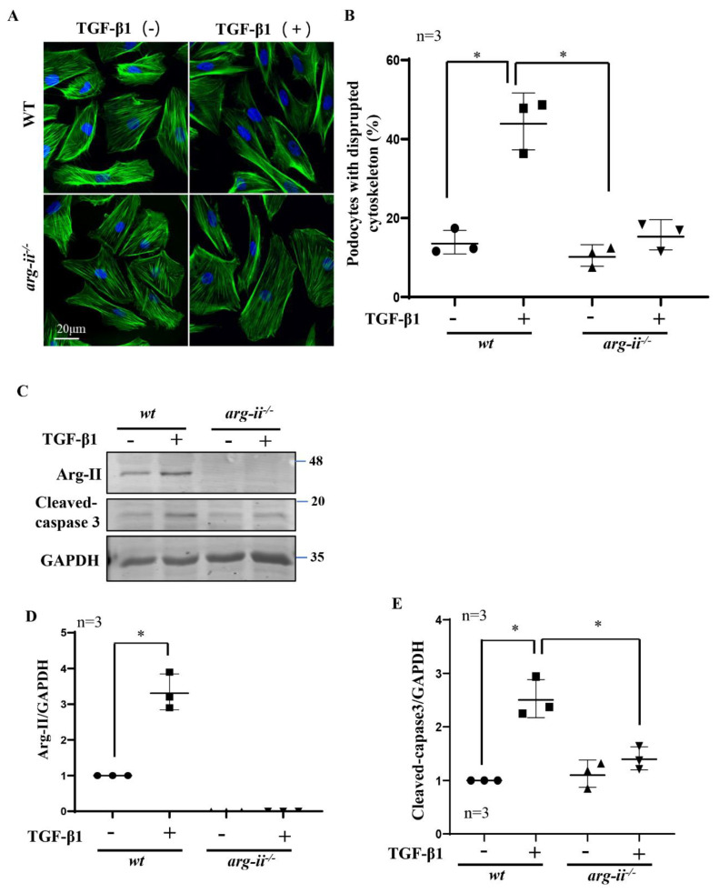
Figure 5.
Arg-ii knockout in podocytes prevents TGF-β1-induced podocyte cytoskeleton disorganization and apoptosis. Podocytes were treated with or without TGF-β1 (10 ng/mL) for 48 h and subjected then to (A) phalloidin staining of cytoskeletal actin fibers (green). The nucleus were stained with DAPI (blue). (B) Quantification of podocytes with disrupted actin cytoskeleton is presented in the graph. (C) Immunoblotting analysis of the protein levels of Arg-II and cleaved-caspase 3 in different groups of podocytes as indicated; GAPDH serves as the loading control. (D,E) Graphs showing quantification of the signals of Arg-II and cleaved-caspase 3, respectively. * p < 0.05 between the indicated groups. n = 3.

