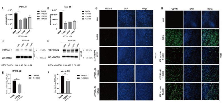
Figure 9.
Monoclonal antibody treatment of ATP1A1 effectively reduces PEDV attachment. (A,B) Treatment with ATP1A1 mAb effectively reduces PEDV RNA abundance. Cells were incubated with serial diluted ATP1A1 mAb at 37 °C for 1 h. Then, the cells were washed with PBS and incubated with PEDV at 0.1 MOI at 4 °C for 1 h. After three washes with PBS, the cells were again incubated with the corresponding mAb in DMEM at 37 °C for 24 h. PEDV RNA abundance was determined by RT-qPCR. Data represent means ± SD from three independent experiments. ***, p < 0.001. (C,D) ATP1A1 mAb treatment significantly decreases PEDV N protein expression. Pretreatment with different folds of diluted ATP1A1 mAb for 1 h, PEDV at 0.1 MOI infected targeted cells for 1 h. Incubation was continued for 48 h in medium containing the corresponding dilution of antibody. Cells were lysed for Western blot analysis. (E,F) ATP1A1 mAb decreases PEDV viral titers. The experimental processing steps were consistent with Figure 8C. At 48 h, virus yields were determined by TCID50 assay with Vero-E6 cells. *, p < 0.05; **, p < 0.01. (G,H) ATP1A1 mAb inhibits PEDV attachment in targeted cells. The experimental processing steps were consistent with Figure 8C. PEDV-infected cells were determined by immunofluorescence staining with anti-PEDV-N mAb (green). Staining of cell nuclei using DAPI (blue) staining solution. Scale bars, 100 μm.

