Figure 3.
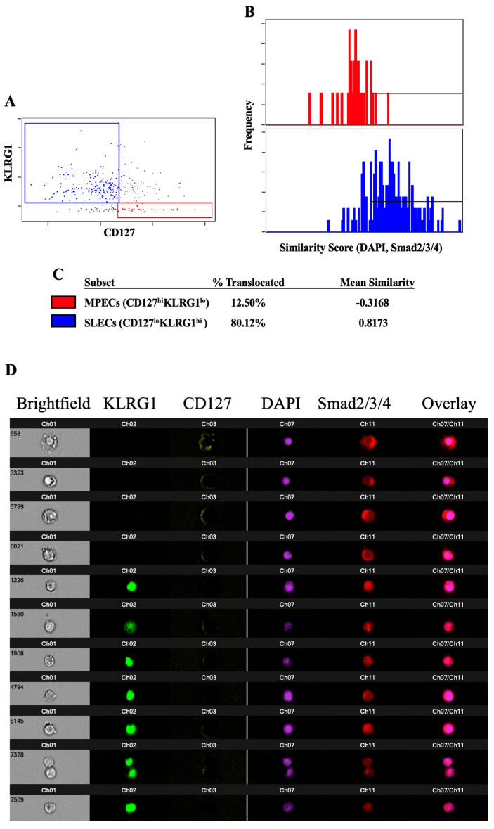
Smad proteins do not efficiently translocate to the nucleus in response to TGFβ signaling in MPECs. Polyclonal CD8 T cells were stimulated with anti-CD3ε and anti-CD28 mAbs for 5 days prior to a 4 h incubation with TGFβ. The cells were then fixed, stained, and analyzed for the nuclear translocation of the Smad2/3/4. (A) Plot of KLRG1 and CD127 demonstrating the gating of MPECs (red) and SLECs (blue). (B) Plot of similarity values between fluorescence indicating the location of Smad2/3/4 and fluorescence indicating the location of the nucleus (DAPI) in MPECs (red) and SLECs (blue). Horizontal line indicates the similarity values that are consistent with the Smad translocation to the nucleus. (C) Calculated percentage of MPECs and SLECs in which Smad2/3/4 have translocated to the nucleus, according to the gate shown in (B), and the mean similarity value for each population. (D) Representative images showing the intensity and location of KLRG1, CD127, the nucleus, Smad2/3/4, and an overlay of the nuclear and Smad images. The data shown are representative of at least three individual experiments with similar results.

