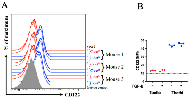
Figure 5.
CD122 expression is not significantly affected by TGFβ signaling. Endogenous MPECs and SLECs were incubated with or without 20 ng/mL TGFβ for 2 h prior to the analysis of CD122 expression by flow cytometry (A). Gray-filled histogram represents isotype control. T-bethi SLECs and T-betlo MPECs from individual mice are represented by blue and red histograms, respectively. (B) CD122 MFI are compared between groups; p > 0.05. The experiment was conducted using three mice per group and repeated twice.

