Figure 3.
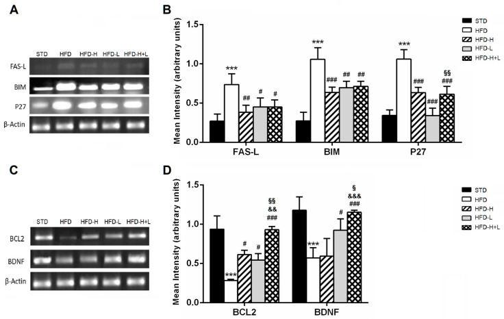
Apoptosis. (A) result of the RT-PCR and (B) mRNA levels of pro-apoptotic genes: FAS-L, BIM and P27 in the mouse brain of different groups; (C) Representative image of the RT-PCR results and (D) mRNA levels of survival genes: BCL2 and BDNF in the mouse brain of different groups. Data are mean values ± S.E.M. (n = 8/group). *** p < 0.001 vs. STD mice; # p < 0.05, ## p < 0.01, ### p < 0.001 vs. HFD mice; && p < 0.01, &&& p < 0.001 vs. HFD-H mice; § p < 0.05, §§ p < 0.01 vs. HFD-L mice.

