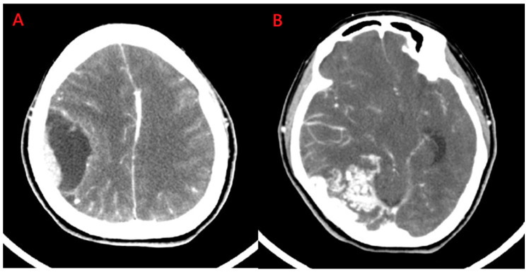
Figure 1.
Computed tomography of brain. (A) The lesion with perifocal edema was seen in the right temporo-parietal area; (B) Another lesion with focal heterogeneous enhancement and a surrounding multilinear contrast structure with a lower density and calcified spots was noted in the right parieto-occipital region.

