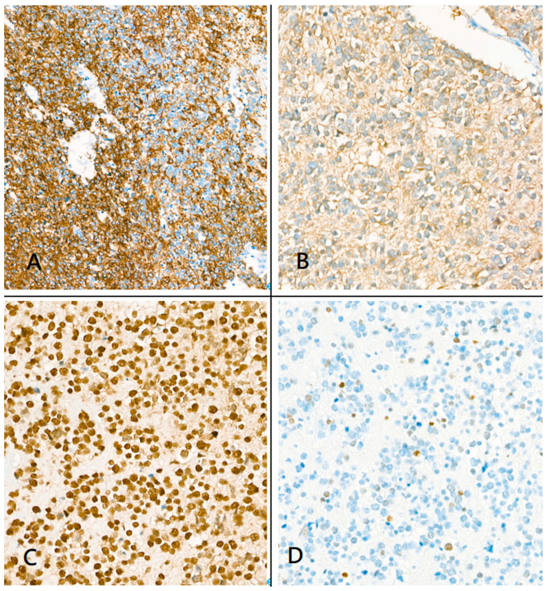
Figure 6.
Immunohistochemical staining. (A) Immunohistochemistry showed that the tumor was positive for glial fibrillary acidic protein; (B) Isocitrate dehydrogenase-1-positive staining; (C) Alpha-thalassemia/intellectual disability X-linked-positive staining; (D) p53 staining showed a wild-type pattern.

