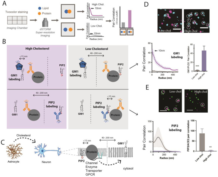
Figure 2.
Strategy for detecting a protein moving between GM1 to PIP2 clusters. (A) Labeling scheme and data analysis. Cells grown in 8-well imaging chambers are stained for a lipid and a protein and imaged with super-resolution. Hypothetical pair correlation data (proximity measurement) is shown. High pair correlation at short distances (e.g., 10 nm) indicates an association. (B) Strategy for establishing cholesterol-dependent localization. Each lipid compartment is labeled pairwise with protein in either high or low cholesterol. Typically, GM1 clusters and proteins are fluorescently labeled with Cholera Toxin B or antibody. (C) Model for CRP regulation in a neuron by astrocyte cholesterol. Astrocytes produce cholesterol which is transported to neurons with apolipoprotein E (ApoE). The neuron takes up the cholesterol into the plasma membrane, where it sequesters proteins with ordered GM1 lipids. (D) Pair correlation data of phospholipase D2 leaving a lipid raft after treating cells to remove cholesterol. Data adapted from Pavel et al., PNAS 2020 [40]. (E) Pair correlation data of angiotensin-converting enzyme 2 (ACE2) moving to PIP2 domains after cholesterol removal. Adapted from Wang et al., 2020 [6]. ** p < 0.01 and *** p < 0.001.

