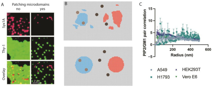
Figure 5.
Antibody patching. (A) antibody patching in unfixed cells. Patches can be seen without super-resolution imaging. Adapted from Lang et al., 2001 with permission [107]. (B) Depiction of antibody patching at nanoscopic levels. In the top panel, there are multiple patches of different sizes. In the bottom panel, clustering helps create lipid domains of more uniform size which is important to the cluster analysis. In light blue, the clustering pulls together proteins more associated with the lipid domains. In red, the clustering separates proteins less associated with the lipid domains. (C) Pair correlation of PIP2 with GM1 in multiple cell types. Pair correlation is very low, suggesting PIP2 and GM1 lipids are separate. A549 are lung epithelial, Human embryonic kidney (HEK293), Vero E6 monkey kidney, and H1793 lung epithelial. Data adapted from Yuan et al., 2022 with permission [7].

