Figure 1.
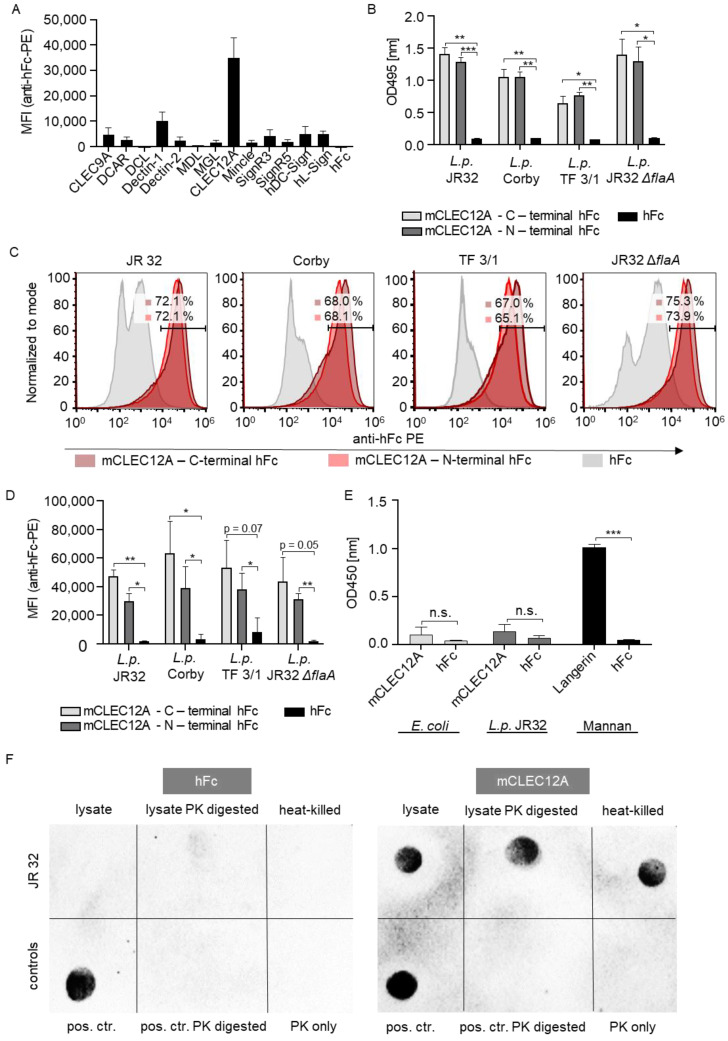
The murine CLEC12A-hFc fusion protein recognizes L. pneumophila (A) Flow cytometry-based binding study of a comprehensive CLR-Fc fusion protein library to L. pneumophila wild-type strain JR32. A PE-conjugated anti-hFc antibody was used for CLR detection. The results are presented as the mean fluorescence intensities (MFI). (B) ELISA-based binding study using the L. pneumophila strains JR32, Corby, TF 3/1 and JR32 ΔflaA. C-terminal hFc (N-CTLD-hFc-C) and N-terminal hFc (N-hFc-CTLD-C) murine CLEC12A fusion proteins were incubated with the respective strains, and binding was detected using an anti-hFc HRP antibody. (C,D) Flow cytometry-based binding study using the L. pneumophila strains JR32, Corby, TF 3/1 and JR32 ΔflaA. The binding study was performed with C-terminal hFc (N-CTLD-hFc-C) and N-terminal hFc (N-hFc-CTLD-C) murine CLEC12A fusion proteins. The detection was performed using a PE-conjugated anti-hFc antibody. (C) Representative histograms of flow cytometry-based binding studies are shown. Values within the histograms show the percentage of L. pneumophila strains that were positive for a PE-conjugated anti-hFc antibody. The first values represent the percentages of the C-terminal hFc murine CLEC12A fusion proteins binding to the respective L. pneumophila strain; the second values represent the percentages of the N-terminal hFc murine CLEC12A fusion protein. (D) The results of the flow cytometry-based binding studies are presented as the mean fluorescence intensities (MFI). (E) ELISA-based binding study using LPS isolated from E. coli and L. pneumophila JR32. The binding of the CLR Langerin to its ligand (mannan) was included as a positive control. (F) Representative dot blot (of 3 independent experiments) using L. pneumophila JR32. The L. pneumophila JR32 samples were pipetted on the upper row as bacterial lysate (“lysate”), proteinase K-digested lysate (“lysate PK digested”) and heat-killed untreated L. pneumophila (“heat-killed”) (from left to right). On the bottom, the following controls were pipetted: hFc as a positive control (“pos. ctr.”), proteinase K-digested hFc (“pos. ctr. PK digested”) and proteinase K only (“PK only”) (from left to right). The left membrane was incubated with hFc (“hFc”) as a control; the right membrane was incubated with murine CLEC12A—C-terminal hFc (“mCLEC12A”). The detection was performed using an anti-hFc HRP antibody. (A–E) All data are shown as the mean ± SD (n = 3). Data were analyzed using a paired Student’s t-test. Asterisks indicate significant differences (n.s. = not significant, * p < 0.05, ** p < 0.01, *** p < 0.001).

