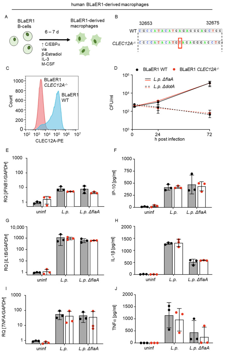
Figure 3.
CLEC12A does not affect the replication of L. pneumophila or the production of proinflammatory cytokines and type I IFNs by BLaER1-derived human macrophages. (A) BLaER1 cells were transdifferentiated into BLaER1-derived macrophages by stimulation of the transcription factor C/EBPα with β-estradiol, IL-3 and M-CSF for 5 to 7 days. (B) BLaER1 CLEC12A−/− cells were generated by introducing a frameshift of one base into CLEC12A (see red box) by CRISPR/Cas9. (C) The loss of CLEC12A in BLaER1 cells was confirmed by flow cytometry, using an anti-CLEC12A-PE labeled antibody. (D) The replication of L. pneumophila (L.p.) ΔflaA and ΔdotA in BLaER1 WT and CLEC12A−/− cells was assessed by infecting cells at MOI 0.1 and evaluating CFUs at 2, 24 and 72 h after infection. BLaER1 WT and CLEC12A−/− cells were infected with L.p. WT and ΔflaA at MOI 10. The expression levels of IFNB1 (E), IL1B (G) and TNFA (I) were measured after 8 h by qRT-PCR and compared with uninfected controls. Data are shown as the relative quantification (RQ) of the target mRNAs relative to GAPDH. (F,H,J) Production of IP-10 (CXCL10) (F), IL-1β (H) and TNFα (J) was measured in supernatants of the infected BLaER1 WT and CLEC12A−/− cells after 18 h. (D–J) All data represent the mean ± SD of 3 independent experiments carried out in triplicate. Differences were assessed using multiple paired t-tests. Comparisons with p < 0.05 were considered significant.

