
FIG. 1.
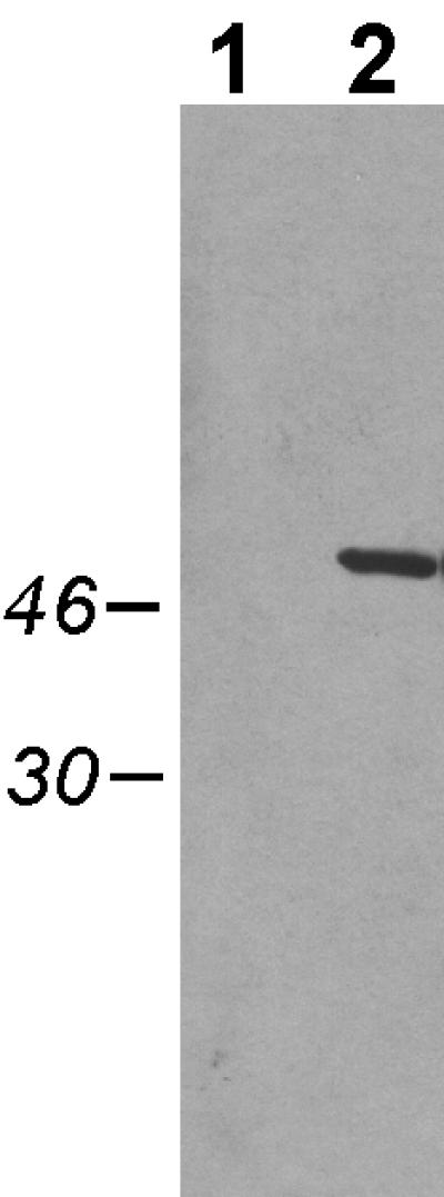
Expression of TadA-T7 in A. actinomycetemcomitans visualized by Western blot analysis. Cells were grown overnight in the presence of 0.1 mM IPTG, pelleted, and then resuspended in SDS loading dye, boiled, and separated by SDS-PAGE through a 12% polyacrylamide gel, as described in Materials and Methods. Lane 1, strain CU1000N carrying pJAK16 (vector); lane 2, strain CU1000N carrying pSK174 (tadA-T7). Numbers (left), molecular masses of relevant markers in kilodaltons.