Figure 1.
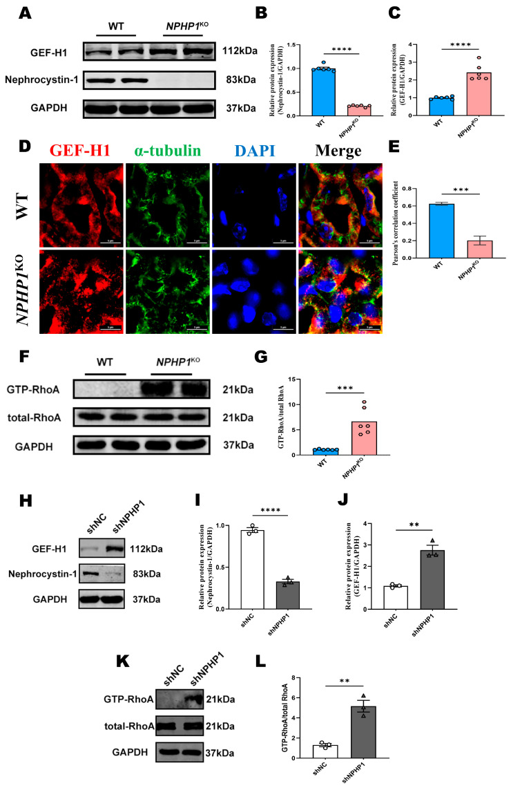
GEF-H1/RhoA pathway was activated in the renal tissue of NPHP1KO mice and NPHP1KD HK2 cells. (A–C): The expression of GEF-H1 was increased in the kidneys of NPHP1KO mice compared with the wild-type (WT) mice by Western blot (A) and densitometry analysis (B,C), (n = 6 mice/group). Nephrocystin-1 was the protein encoded by NPHP1 gene. (D): Intracellular spatial distribution of microtubules and GEF-H1 in WT and NPHP1KO mice; In NPHP1KO mice, GEF-H1 was more concentrated on the luminal side of the tubules (red), but the expression and intracellular distribution of microtubules remained unchanged (green) (Immunofluorescence, scale bar: 5 μm). (E): Pearson’s correlation coefficients showing the degree of colocalization of microtubules and GEF-H1 under different conditions (n = 6 mice/group). (F,G): GTP-RhoA were significantly upregulated in the kidney tissue of NPHP1KO mice (F): Western blot, (G): Densitometry analysis, (n = 6 mice/group). (H–J): The expression of GEF-H1 was increased in NPHP1KD HK2 cells (shNPHP1) compared with control cells (shNC) (H): Western blot, (I,J): Densitometry analysis, (n = 3). (K–L): GTP-RhoA was significantly upregulated in NPHP1KD HK2 cells (shNPHP1) (K): Western blot, (L): Densitometry analysis, n = 3). Data represent the mean ± SEM. ** p < 0.01; *** p < 0.001; **** p < 0.0001; ns, no significance.

