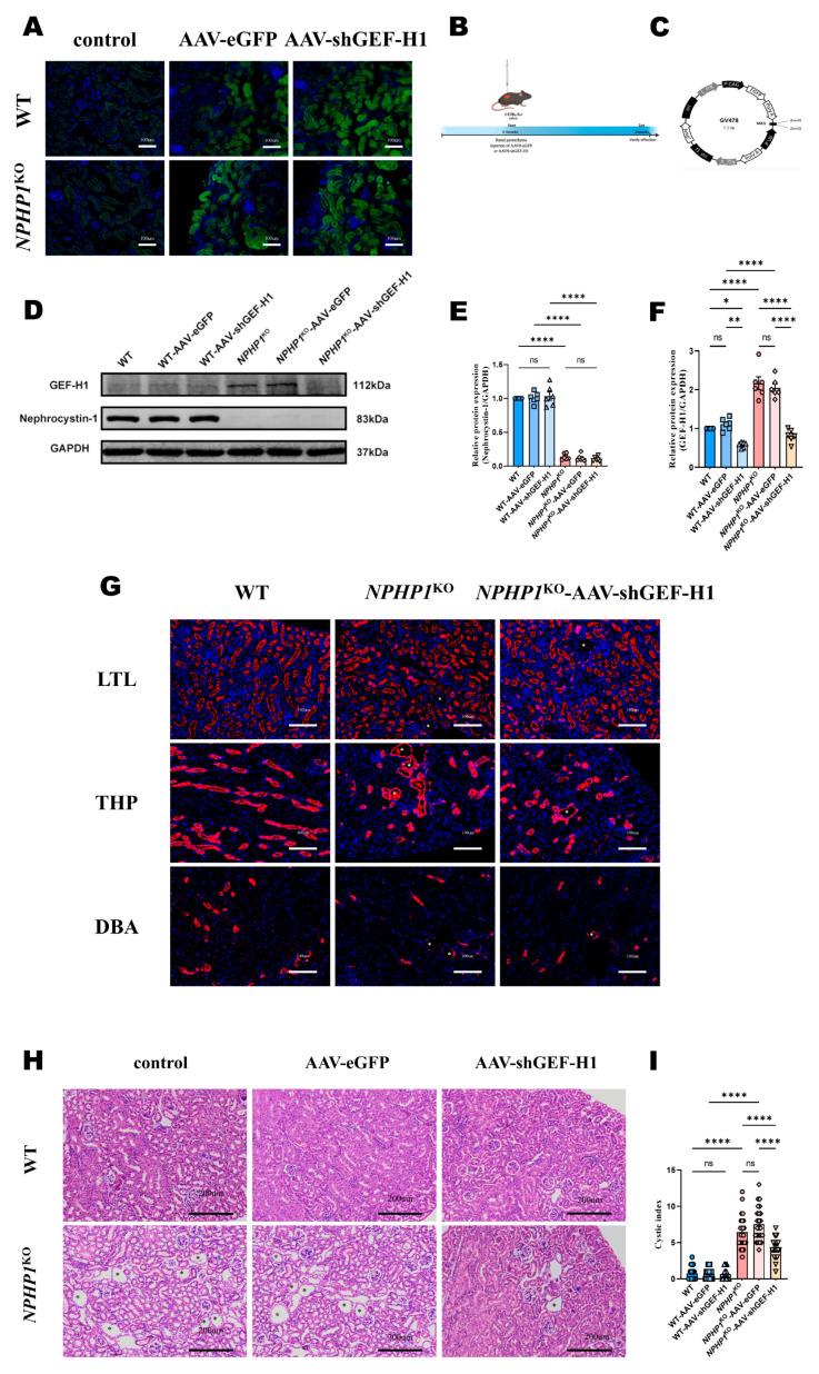
Figure 2.
GEF-H1 knockdown reduced renal cyst formation in NPHP1KO mice compared to WT mice. (A): Immunofluorescence analysis of adeno-associated virus (AAV) transfection efficiency. AAV-eGFP and AAV-shGEF-H1 transduced kidneys expressed eGFP (green). The sections were counterstained with DAPI (blue). (Light microscope, scale bar = 100 μm). (B): Time course of AAV9-mediated GEF-H1 knockdown. (C): Structural diagram of the AAV vector GV478. (D–F): The expression of GEF-H1 and nephrocystin-1 levels in mouse kidney tissue were evaluated by Western blot (D) and densitometry analysis. (E,F): Densitometry analysis; n = 6 mice/group). The increased GEF-H1 level of NPHP1KO mice was then decreased after being transfected with AAV-shGEF-H1. (G): Staining of the kidney with lotus tetragonolobus lectin (LTL+ means proximal tubules; red), Tamm-Horsfall protein (THP+ means distal convoluted tubules; red), and Dolichus biflorus agglutinin (DBA+ means collecting ducts; red). Scale bar = 100 μm. (H): Mouse kidney histopathological changes (hematoxylin-eosin (HE) staining, scale bar = 200 μm). Asterisks indicate the cysts. Cyst formation and renal tubular dilatation in NPHP1KO mice were alleviated after GEF-H1 knockdown. (I): Quantification of the cystic index in mouse kidney specimens (dots represent the number of cysts from 200× magnified images (4–5 images/mouse) (n = 6 mice/group). Data represent the mean ± SEM. * p < 0.05; ** p < 0.01; **** p < 0.0001; ns, no significance.

