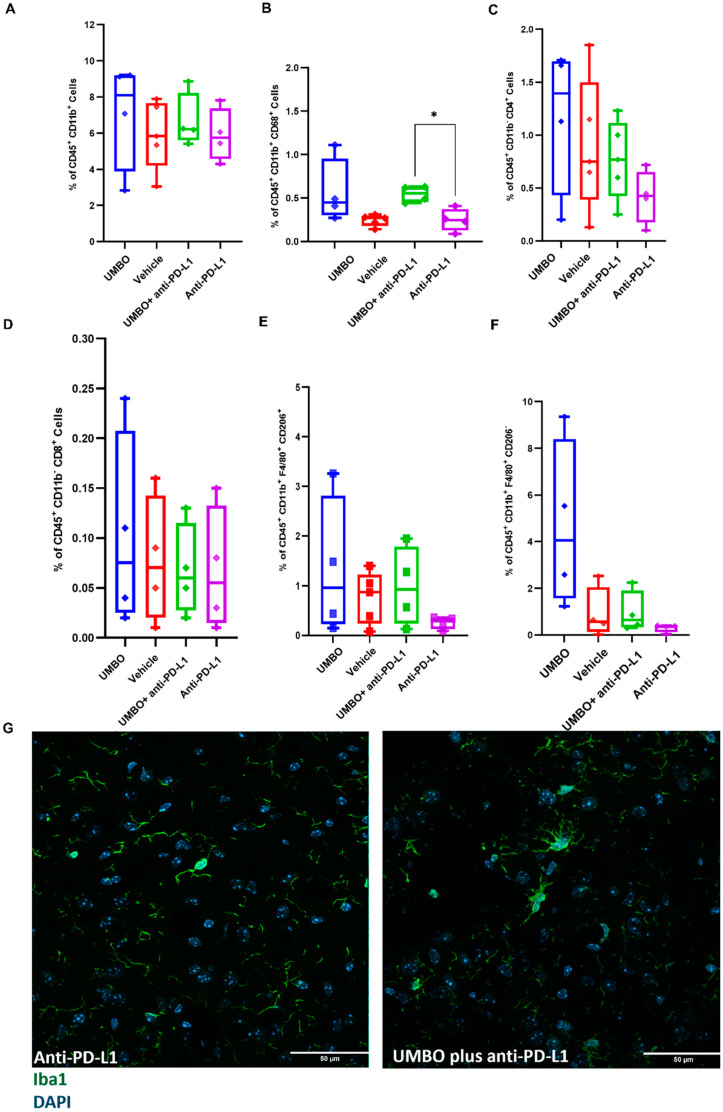
Figure 5.
UMBO plus anti-PD-L1 activates microglia and modulates microglial phenotype. (Panel A): Flow cytometry analysis of the percentage of total microglia in all different groups (n = 4–5). (Panel B): UMBO plus anti-PD-L1 significantly enhanced (* p < 0.05) the percentage of CD68+ cells than an-ti-PD-L1 alone. (Panel C,D): UMBO plus anti-PD-L1 did not influence CD4+ and CD8+ T-lymphocytes percentages compared to other groups. (Panel E,F): No significant difference in CD206+ and CD206− macrophages in all groups. UMBO plus anti-PD-L1 did not modulate macrophages’ expression. (Panel G): Green: Iba1 microglia/macrophages Blue: DAPI nuclear staining; microglia staining in anti-PD-L1 plus UMBO (right photos) treated group confirm a phenotype of activated microglia.

