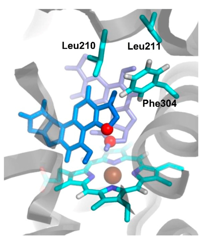
Figure 18.
Binding mode of doubly ligated AFB1-CYP3A4 complex obtained by docking studies. Amino-acid residues Leu210, Leu211, Phe304, and the heme group are shown in sticks. The first AFB1 molecule docked is shown in violet and the second AFB1 molecule is shown in blue. The atoms are colored as follow: carbon in cyan, oxygen in red, nitrogen in blue, hydrogen in white, and iron in brown. Adapted from Ref. [61].

