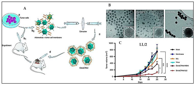
Figure 11.
(A) Tumour cells (a) were cultured and engrafted into mouse model (b1). (b2) The cell membrane was extracted and mixed with an oncolytic adenovirus serotype 5, with a 24-base-pairs deletion, carrying -CpG islands (i.e., A5-Δ24-CpG). (c) The virus was wrapped with the cell mem brane using the process of extrusion to obtain ExtraCRAd. (d) The established tumours were treated with multiple intratumoral injections of ExtraCRAd. (B) Cryo-transmission electron microscopy (TEM) images of virus, lipid cancer membrane vesicles, and ExtraCRAd (C) Median tumour growth [138]. The results were analyzed with a two-way ANOVA, and Dunnet’s post-test comparison, and the levels of significance were * p < 0.05, *** p < 0.001, **** p < 0.0001. Copyright CC BY 4.0.

