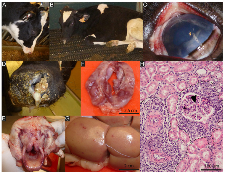
Figure 3.
Typical lesions of bovine WD-MCF induced by AlHV-1 infection. (A,B) Calves showing typical head and eye form of WD-MCF, with apathy, ptyalism, mucopurulent discharge, and muzzle crusting. (C) Corneal opacity and mucosal petechia. (D) Detail of the muzzle illustrating the presence of adherent fibrino-necrotic material that partially occludes the nasal orifices. (E) Ulcerative and necrotic pharyngitis and laryngitis. (F) Longitudinal section through an inguinal lymph node. Note the loss of normal tissue structure of the organ and necrotic appearance. (G) Irregularly distributed white to reddish foci of a few millimeters in diameter deform the surface of the kidney. (H) Hematoxylin and eosin-stained kidney section highlighting the accumulation of lymphoblastic cells around small arterioles (dashed curves). Arrowhead indicates renal corpuscle.

