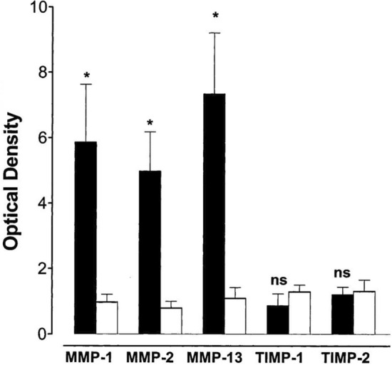
Fig. 3.

Protein expression of MMPs and their TIMPs in extracts of skin samples from patients with stasis dermatitis. Data were taken from Western blots with monoclonal antibodies against MMP-1, MMP-2, MMP-13, TIMP-1, and TIMP-2 in stasis dermatitis and control skin lesions. Densitometric evaluations of immunoreactive blots for MMP-1, MMP-2, MMP-13, TIMP-1, TIMP-2 from stasis dermatitis (black bars) and healthy skin (white bars) are shown. Data are mean ±SEM; n = 10 for stasis dermatitis and n = 4 for control. The significance of difference was determined by an unpaired Student t test and is indicated in each mapped group. Figure used with permission from Herouy et al. [46] J Dermatol Sciences, 2001. MMP matrix metalloproteinase, ns not significant, SEM standard error of the mean, TIMP tissue inhibitor of metalloproteinase. *p < 0.05
