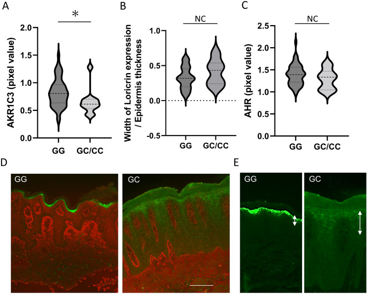
Figure 3.
Results of immunohistochemistry. Immunostaining for AKR1C3 and loricrin was performed on formalin-fixed paraffin-embedded tissues of psoriasis lesions from 62 patients. (A) AKR1C3 expression in the epidermis was significantly lower in psoriasis lesional skin samples from patients with rs12529 G > C and rs12387 A > G than in patients with rs12529 G/G and rs12387 A/A (p = 0.0434, Student’s t-test). (B) Wide loricrin expression due to abnormal early expression was observed in the epidermis from patients with rs12529 G > C and rs12387 A > G SNPs. (C) AHR expression did not differ depending on the variant. (D) Representative immunostaining images of AKR1C3 (red) and loricrin (green) staining. (E) Ectopic expression of loricrin in the spinous layer in patients with the rs12529 G > C SNP.

