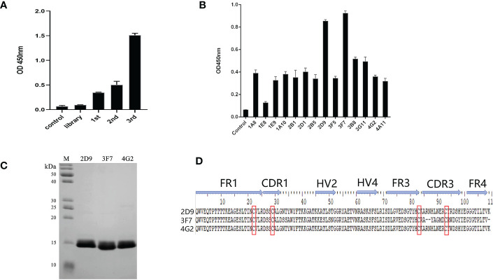
Figure 3.
Selection of anti-OGT VNARs. (A) Detecting the titer of phage single domain antibody library in each round of panning; (B) Detection of specific bacteriophage by monoclonal ELISA; (C) Characterization and analysis of molecular weight of 2D9,3F7 and 4G2; (D) Amino acid sequence alignment of the three anti-OGT VNARs. FR is framework region; CDR is complementarity-determining region; HV is hypervariable region. Canonical Cys residues are enclosed in red.

