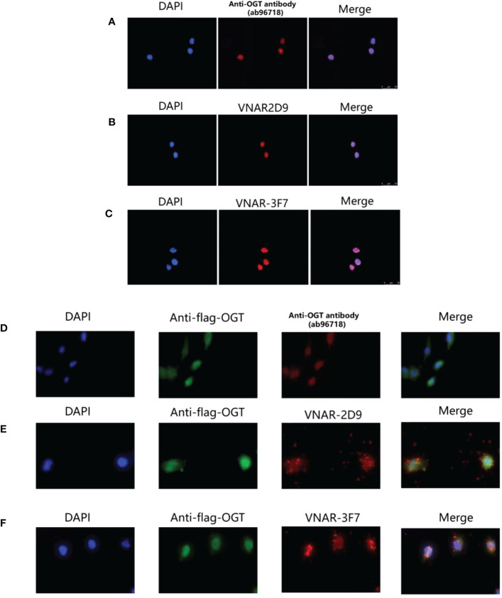
Figure 8.
Cytosolic and cytoplasmic localization of OGT. (A) Binding of anti-OGT/O-linked N-acetylglucosaminyltransferase (ab96718); (B) 2D9; (C) 3F7 in wild-type H1299 cells, respectively. Immunofluorescence images representing OGT (Red) and DAPI (Blue) were overlaid (Merge) to show localization within the nucleus. (D–F). Commercial anti-OGT antibody (D), 2D9 (E), and 3F7 (F) were each co-localized with p3×flag-ncOGT transfected H1299 cells. Immunofluorescence images of OGT represented in green, the three antibodies represented in Red and DAPI Blue were overlaid (Merge) to show co-localization.

