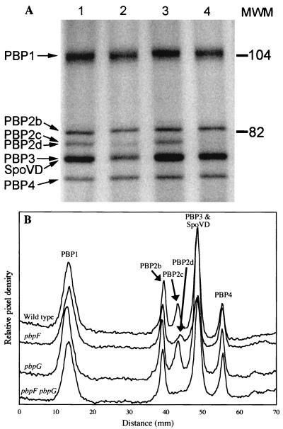
FIG. 1.
PBP profiles of wild-type, pbpF mutant, and pbpG mutant strains. Membranes were purified from cultures at the fourth hour of sporulation. (A) Membranes were incubated with 125I-labeled penicillin X, proteins were separated on an SDS–7.5% PAGE gel, and PBPs were detected using a phosphorimager. Lane 1, wild type; lane 2, pbpF; lane 3, pbpG; lane 4, pbpF pbpG. Calibrated molecular mass standards (MWM; in kilodaltons) were Bio-Rad low-range-prestained SDS-PAGE standards. PBP2a decreases dramatically during sporulation (7) and is not visible on this gel. (B) Histogram of PBP band intensities produced by integrating signal strength within columns that covered 90% of each lane's width. PBPs are numbered as previously described (5, 23).

