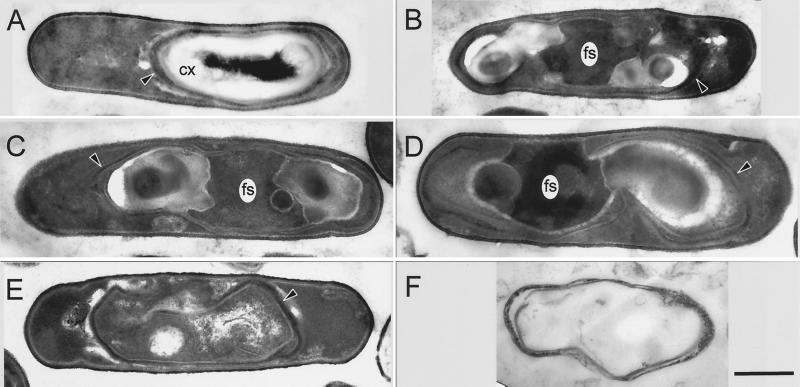
FIG. 3.
Thin-section electron microscopy of sporulating mutant cells. Cells of pbpG (A to C) or pbpG pbpF (D to F) strains were sporulated, harvested at hour 7 (A to D) or 24 (E and F), and prepared for electron microscopy. The majority of pbpG cells had an appearance (A) similar to that of the wild type. In a minority of the pbpG cells, masses, presumed to be spore PG, do not completely surround the forespore (B and C). The majority of pbpF pbpG cells had this appearance (D). (E) A pbpF pbpG double mutant cell at the 24th hour of sporulation, in which the masses on either side of the forespore have disappeared. (F) Empty spore coat structure released by lysis of a pbpF pbpG double-mutant cell at the 24th hour of sporulation. CX, cortex; fs, forespore cytoplasm. Arrowheads, spore coat structures. Bar (F), 500 nm (all panels have the same magnification).

