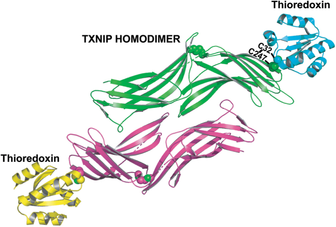
FIG. 2.

The TXNIP-thioredoxin interaction. Crystal structure of the TXNIP dimer-thioredoxin complex, resolution 2.7 Å, balls refer to bonding cysteine residues, green and pink are beta sheets and disoriented regions of TXNIP, and yellow and cyan are alpha helices and beta sheets of thioredoxin. Created using PyMOL Molecular Graphics System.
