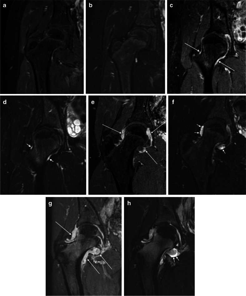
Fig. 2.
Overall degree of inflammation in right-hip MRIs in four pairs of images from four children with juvenile idiopathic arthritis (JIA) across different levels of severity. All images are demonstrated using coronal post-contrast three-dimensional (3-D) gradient echo with fat saturation (a, c, e, g) and coronal fat-saturated T2-weighted turbo spin-echo (b, d, f, h) MRI sequences. a, b No inflammation (score 0) in a 12-year-old boy. c, d Mild synovial thickening with moderate increase in post-contrast enhancement (long arrows) and sliver of effusion (short arrows) (score 1) in a 16-year-old girl. e, f Moderate synovial thickening with moderately increased post-contrast enhancement (long arrows) and mild effusion (short arrows) (score 2) in a 15-year-old boy. g, h Severe synovial thickening and increased post-contrast enhancement, more evident at the medial aspect of the joint (long arrows), with mild/moderate effusion (short arrows) (score 3) in a 17-year-old girl

