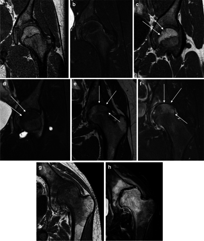
Fig. 3.
Femoral head bone marrow edema demonstrated on MRI of the left hip in four pairs of images from four children with juvenile idiopathic arthritis (JIA) across different severity levels. Bone marrow edema was defined as hypointense areas on T1-W sequences with corresponding hyperintense areas on fat-saturated T2-W sequences in the bone marrow. All images are demonstrated using coronal three-dimensional (3-D) T1-weighted turbo spin-echo (a, c, e, g) and fat-saturated T2-weighted turbo spin echo (b, d, f, h) MRI sequences. a, b No visible bone marrow edema (score 0) in a 13-year-old boy. c, d Two focal areas of bone marrow edema (less than 33% of the bone volume, arrows) (score 1) in a 11-year-old girl. e, g Large area of bone marrow edema (between 33 and 66% of the bone volume, arrows) (score 2) in a 14-year-old girl. g, h Widespread bone marrow edema (almost 100% of the bone volume) (score 3) in a 15-year-old boy

