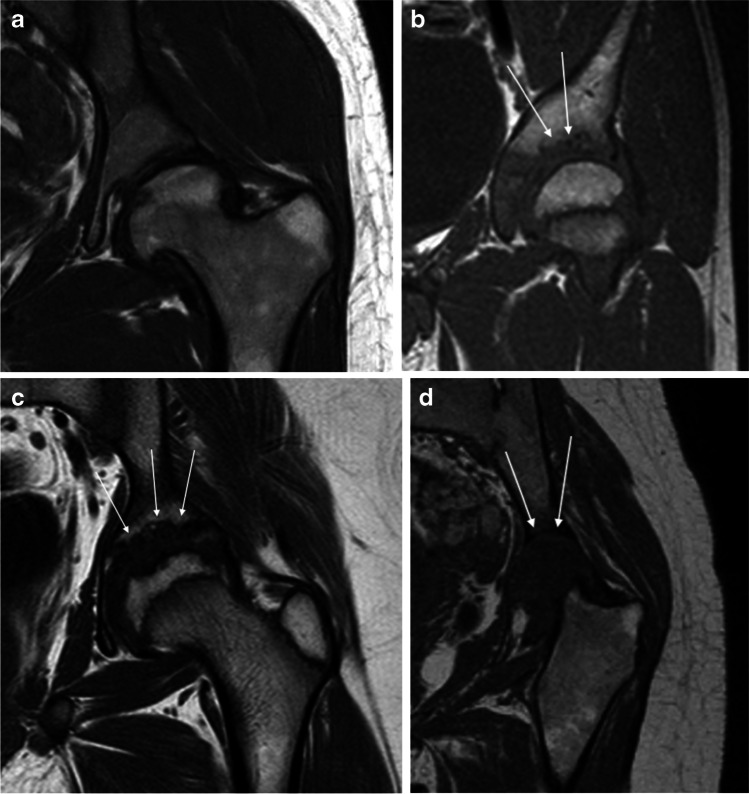
Fig. 4.
Erosions demonstrated on MRI at the left acetabulum as shown on coronal three-dimensional (3-D) T1-weighted turbo spin-echo sequences in patients with juvenile idiopathic arthritis (JIA). a No visible acetabular erosions (score 0) in a 20-year-old woman. b Some erosions on the superior aspect of the acetabulum (< 33% of the surface, arrows) (score 1) in a 18-year-old man. c Multiple acetabular erosions (between 34% and 66% of the surface, arrows) (score 2) in a 13-year-old boy. d Erosive changes of the whole acetabular surface (arrows) (score 3) in a 19-year-old woman with complete destruction of the femoral head (arrows)

