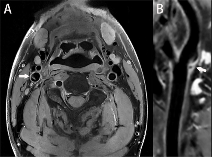
FIGURE 4.
(A) A high-resolution magnetic resonance T1-weighted imaging (T1WI) axial image shows a thin, membrane-like structure protruding from the wall of the internal carotid artery to the lumen; furthermore, this structure separates the lumen (arrow). (B) A T1WI-enhanced sagittal image shows a locally enhanced, high signal on the carotid web (arrow).

