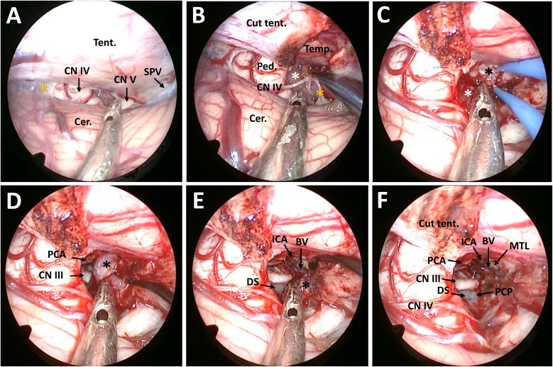
Figure 2.
Surgical procedure and nuances of the EF-SCITA approach for resection of posterior clinoid meningioma. (A) Endoscopic explorations in the lateral supracerebellar infratentorial space and the exposure of CN IV after opening the arachnoid in the ambient cistern. (B) The exposure of the infratentorial part of the tumor in the ambient cistern after incision of the tentorium cerebelli. (C) The exposure of the supratentorial tumor after up-elevating the tentorium cerebelli. (D) The exposure of the PCA and CN III as well as the supratentorial tumor after debulking of the infratentorial tumor. (E) Dissection of the supratentorial tumor from the ICA and the initial part of BV and the exposure of DS during tumor resection. (F) The exposure of PCP after total tumor removal. BV, basal vein of Rosenthal (the initial part); Cer., cerebellum; CN III, oculomotor nerve; CN IV, trochlear nerve; CN V, trigeminal nerve; DS, dorsum sellae; ICA, internal carotid artery; MTL, medial temporal lobe; PCA, posterior cerebral artery; PCP, posterior clinoid process; Ped., pedunculus cerebri; PS, pituitary stalk; SPV, superior petrosal vein; Tent., tentorium cerebelli. The black asterisks indicate the supratentorial part of the tumor, the white asterisk denotes the infratentorial part of the tumor, and the yellow asterisks indicate the arachnoid in the ambient cistern.

