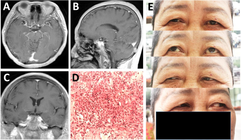
Figure 3.
Radiological and pathological evaluation of the lesion postoperatively. (A-C) Postoperative post-contrast axial (A), sagittal (B), and coronal (C) MRI scans performed one month after surgery showing total removal of the lesion. (D) Photomicrograph of hematoxylin-eosin staining showing typical features of meningothelial meningioma. (E) Physical examinations at one month follow-up indicating no limitation in eye movements.

