



Abstract
Erythropoietic protoporphyria (EPP) and X-linked protoporphyria (XLP) are rare genetic photodermatoses. Limited expertise with these disorders among physicians leads to diagnostic delays. Here, we present evidence-based consensus guidelines for the diagnosis, monitoring, and management of EPP and XLP. A systematic literature review was conducted, and reviewed among sub-committees of experts, divided by topic. Consensus on guidelines was reached within each sub-committee and then among all members of the committee. The appropriate biochemical and genetic testing to establish the diagnosis is reviewed in addition to the interpretation of results. Prevention of symptoms, management of acute phototoxicity, and pharmacologic and non-pharmacologic treatment options are discussed. The importance of ongoing monitoring for liver disease, iron deficiency, and vitamin D deficiency is discussed with management guidance. Finally, management of pregnancy, surgery, and the safety of other therapies are summarized. We emphasize that these are multisystemic disorders that require longitudinal monitoring. These guidelines provide a structure for evidence-based diagnosis and management for practicing physicians. Early diagnosis and management of these disorders are essential, particularly given the availability of new and emerging therapies.
Keywords: erythropoietic protoporphyria, X-linked protoporphyria, protoporphyria, EPP, XLP, guidelines, evidence-based, consensus, diagnosis, management, cutaneous porphyria, photodermatoses
Introduction
Erythropoietic protoporphyria (EPP) and X-linked protoporphyria (XLP) are rare genetic disorders of the heme biosynthetic pathway characterized by the accumulation of protoporphyrin in erythrocytes, plasma, and the biliary system.1 Although indistinguishable clinically, EPP results from loss-of-function variants in ferrochelatase (FECH), the last enzyme of heme biosynthesis, and XLP results from gain-of-function variants of aminolevulinate synthase 2 (ALAS2), the first enzyme of heme biosynthesis (Figure 1).2 In both, protoporphyrin released from erythroid cells into the vascular endothelium and surrounding tissues is activated by visible light, triggering oxidative stress and inflammation.3 Patients are primarily sensitive to visible light in the blue range.1 Both disorders present in childhood with severe, non-blistering phototoxicity preceded by characteristic prodromal symptoms, often including tingling, burning, or itching that serve as warning signals to avoid sun exposure.1 Continued exposure leads to severe phototoxic pain that can be associated with erythema and swelling of sun-exposed skin, primarily the dorsum of the hands and face (Figures 2, S1). The pain is usually not responsive to analgesics, and recovery may take several days. Recurrent phototoxic episodes result in a conditioned behavior of sunlight avoidance and decreased quality of life.4,5 In addition to phototoxic episodes, these disorders have multisystemic manifestations including hepatopathy, iron deficiency anemia, and vitamin D deficiency, which are often under-recognized.1
Figure 1. Molecular Pathophysiology of the Protoporphyrias.

The heme biosynthetic pathway requires eight enzymatic steps. Gain-of-function variants in ALAS2 result in X-linked protoporphyria (XLP), and loss-of-functions variants in FECH result in erythropoietic protoporphyria (EPP). In both, metal-free protoporphyrin IX accumulates in erythroblasts, erythrocytes, the plasma, and the biliary system. Metal-free protoporphyrin IX is photosensitive, particularly to visible light in the blue range, and the light-mediated activation of metal-free protoporphyrin IX produces free radicals that damage the surrounding tissues. ALAS2, aminolevulinate synthase 2; FECH, ferrochelatase; ALAD, delta-aminolevulinic acid dehydratase; PBGD, porphobilinogen dehydratase; UROS, uroporphyrinogen III synthase; UROD, uroporphyrinogen III decarboxylase; CPOX, coproporphyrinogen-III oxidase; PPOX, protoporphyrinogen oxidase
Figure 2.
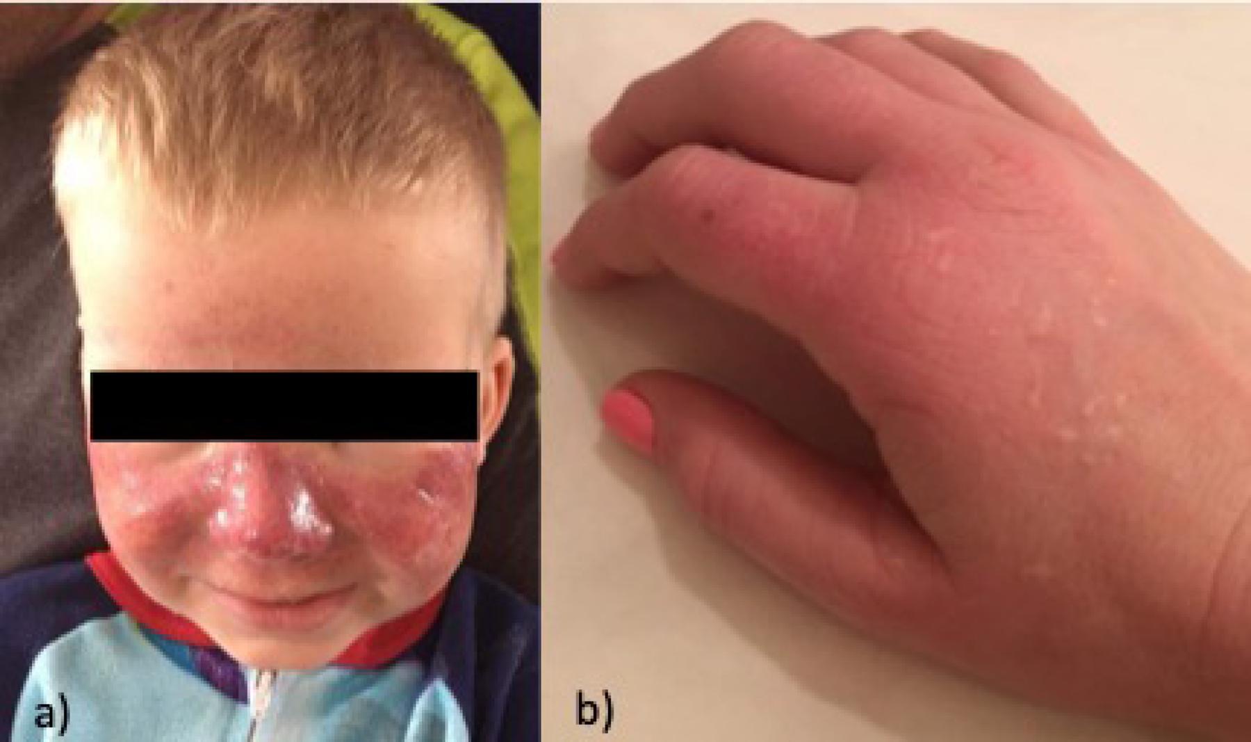
Cutaneous findings in protoporphyria
a) Child with extensive edema of the face with erythema and petechiae
b) Adult patient with erythema and edema during phototoxic episode, with hypopigmented scars and skin thickening present
Of note, in protoporphyria, the skin may have no visible changes during severely painful phototoxic episodes.
The mean diagnostic delay is over a decade, and evidence supports significant underdiagnosis.6,7 The disease prevalence of EPP was estimated to be 1:109,000 in Europe, but more recent data using exome databases show a prevalence approximating 1:17,000 in Caucasians.8,9
There are few specialists with expertise in these rare disorders. Many patients are managed by dermatologists or primary care physicians with limited experience with these disorders and no standardization of care. Because new therapies are now available and more are in development, the diagnosis and identification of patients who may benefit from therapeutic interventions or need closer follow up for long-term complications is particularly important. Here, we provide the first evidence-based consensus guidelines for the diagnosis and management of these photodermatoses.
Scope
These guidelines address the diagnosis, monitoring, and management of patients with EPP and XLP (referred to collectively as protoporphyria in this manuscript) without evidence of liver disease. Management of protoporphyria-mediated hepatopathy and indications for liver or bone marrow transplantation are outside the scope of these guidelines. Multidisciplinary consultation at a center with hematology, hepatology, and liver transplantation expertise in protoporphyria is recommended for patients with progressive liver disease.
Methods
A systematic review of the literature was performed using best practices from the Cochrane Handbook for Systemic Reviews of Interventions and the Preferred Reporting Items for Systemic Reviews and Meta-Analyses (PRISMA) statement.10,11 The review examined all primary research studies pertaining to diagnosis and management using MESH headings “erythropoietic protoporphyria” and “X-linked protoporphyria,” including randomized controlled trials, cohort studies, case-control studies, case reports, and cross-sectional studies. The search was conducted in the following databases with no language or date restrictions: PubMed, EMBASE, Web of Science, and Clinicaltrials.gov. Duplicates and conference abstracts were removed.
Inclusion criteria were studies related to the diagnosis and management of EPP or XLP and human or animal studies. Exclusion criteria were studies that were not accessible in English, did not pertain to EPP or XLP, conference abstracts, and non-primary research. Two reviewers independently screened titles and abstracts in Covidence using these pre-defined eligibility criteria, and differences in opinions were resolved by discussion and consultation with a third reviewer.12 The full text of the remaining studies was screened for further exclusion (Figure S2).
A writing committee composed of 17 clinicians who were members of the Porphyrias Consortium and with recognized expertise were selected to establish diagnostic and management guidelines.13 The committee included experts in dermatology, hematology, hepatology, genetics, neurology, and internal medicine, and one physician-patient. The committee was divided into the following topic subgroups: diagnosis, dermatology, hepatology, hematology, and miscellaneous. Members of each subgroup reviewed articles relevant to their topic. Subgroups arrived at evidence-based guidelines addressing the important diagnostic and management concerns for that topic. For diagnostic or management concerns with minimal to no evidence (level C evidence), recommendations were determined by consensus within each subgroup followed by the entire writing committee, achieved through a series of small and large group meetings and voting sessions. The word choices of the recommendations were selected to be consistent with the size of treatment effect, indicated by the recommendation class.14
Results
Diagnosis
Total erythrocyte protoporphyrin concentration, including proportions of metal-free and zinc-bound protoporphyrin, is the recommended test for the diagnosis of protoporphyria (Table 1).15–17 The percentage of metal-free protoporphyrin is typically >90% in EPP and ~50–85% in XLP.2 Minor elevations in protoporphyrin (mostly zinc-bound and/or <3x upper limit of normal) is not consistent with a diagnosis of protoporphyria, and further evaluation for other causes such as iron-deficiency anemia or lead intoxication can be beneficial.15–17 Laboratories that can accurately fractionate metal-free and zinc-bound protoporphyrins are recommended to make the diagnosis (Table 1).15–17 Clinicians should avoid laboratories that measure protoporphyrin only by hematofluorometry, which can lead to falsely normal levels of erythrocyte protoporphyrins, and these results are often incorrectly labeled as “free erythrocyte protoporphyrin.” Clinicians should refer to the United Porphyrias Association website for currently recommended laboratories.18 Because exposure of samples to light may cause inaccuracies in protoporphyrin testing, instructions on light-protected specimen handling should be provided to both phlebotomists and patients.
Table 1.
Diagnosis and Monitoring in Protoporphyria
| Primary diagnostic test | Laboratory tests that should be evaluated at least yearly |
|---|---|
|
|
| Follow-up tests to distinguish EPP from XLP | Monitoring for patients on afamelanotide |
|
|
| Tests not recommended for diagnosis | |
| |
| |
| Level of Evidence | |
| Class I, level B: benefit >>> risk, limited published data | |
| Class I, level C: benefit >>> risk, very limited to no published data | |
| Class III no benefit, level B: no benefit, limited published data | |
| Class III no benefit, level C: no benefit, very limited to no published data | |
In patients with protoporphyria, plasma porphyrin levels may be normal or increased. Elevated plasma porphyrins are not diagnostic of EPP and are therefore not recommended as first-line testing.19 Urinary and fecal porphyrins are typically normal in protoporphyria, and testing is not recommended.20,21 Skin biopsies are not indicated for the diagnosis of protoporphyria and should be reserved for cases where other diagnoses are being ruled out.22
If a diagnosis of protoporphyria is established based on significantly elevated protoporphyrin, genetic testing of the FECH and ALAS2 genes is recommended to distinguish between EPP and XLP.2 Approximately 4% of patients will not have a causative pathogenic variant identified even with elevated protoporphyrin.1 The vast majority of patients with EPP are compound heterozygotes for both a rare pathogenic FECH variant and a common low expression FECH allele that is present in ~10% of the Caucasian population (c.315-48T>C), a combination that results in ~30% of normal enzyme activity.2,8 Homozygosity for FECH c.315-48T>C does not cause EPP. 23 Genetic testing in the absence of biochemical testing is not recommended given the possibility of identifying variants of uncertain significance and the small percentage of patients in whom the genetic variant remains undefined. XLP accounts for 2–10% of protoporphyria cases and is caused by gain-of-function variants in exon 11 of ALAS2 and follows an X-linked inheritance pattern.24 Because of the possibility of skewed X-inactivation, disease severity in females can vary from mild to as severe as XLP males.24 Genetic counseling is recommended for couples with a personal or family history of protoporphyria who are planning to have children.
Management of dermatologic symptoms
Pharmacologic management
Although various pharmacologic agents have been studied for EPP, only a few robust clinical studies evaluating efficacy are available.25 Studies of afamelanotide, a subcutaneously administered, long-acting α-melanocyte stimulating hormone analogue demonstrate that it increases the duration of pain-free sun exposure and improves quality of life in adults with EPP.25–29 Afamelanotide binds to the melanocortin-1 receptor (MC1R) and increases eumelanin production from dermal cells and melanocytes, thus increasing pigmentation and providing photoprotection. Melanin is additionally a strong antioxidant thought to inactivate the reactive oxygen species produced during phototoxic reactions.30 Long term follow-up studies reported significant improvement in quality of life and a high rate of therapy continuation.28 Therefore, afamelanotide is recommended for the prevention of phototoxic symptoms in EPP (Table 2).25–29 For patients using afamelanotide, total body skin examinations by a dermatologist are recommended every 6 months. The drug is available in the US and Europe, however access is limited in the US due to the high cost and restricted sites for administration. There is no data on the safety of afamelanotide during pregnancy so this cannot be recommended for pregnant women. Due to insufficient data related to efficacy, the following therapies are not recommended for the prevention of phototoxic symptoms: beta-carotene, cysteine, cimetidine, isoniazid, warfarin, quinacrine, oral zinc, N-acetylcysteine, vitamin C, omega-3 fatty acids, oral adenosine monophosphate, canthaxanthine, terfenadine, inosine, dithiothreitol (DTT) and glycerol, pyridoxine, and hydroxyethylrutosides (Table 2).25,31–52 Although several previous studies have investigated beta carotene in EPP, the evidence shows unclear or no benefit.25,31,51,52 Dersimelagon is a synthetic, orally administered, small molecule agonist of MC1R being tested for the prevention of protoporphyria phototoxicity. A positive Phase 2 clinical trial has been completed showing promising results, and a Phase 3 study is ongoing.53 There are no approved therapies for pediatric patients.
Table 2.
Management of Protoporphyria
| Medical and non-medical management for the prevention of phototoxic symptoms | Therapeutics with no clear benefit for the prevention of phototoxic symptoms |
|---|---|
|
|
| |
Management only to be considered in patients who are not a candidate for afamelanotide:
| |
| |
| Management of phototoxic symptoms |
Therapeutics with no clear benefit for the
treatment of phototoxic symptoms |
|
|
| Iron deficiency in ferrochelatase-deficient EPP | |
If symptomatic from iron deficiency and/or have hemoglobin levels <10g/dL and ferritin <10ug/L:
| |
| Prevention of liver disease | |
| Iron deficiency in XLP |
|
If any iron deficiency:
| |
| |
| Pregnancy management | |
| |
| Levels of Evidence | |
| Class I, level B: benefit >>> risk, limited published data | |
| Class I, level C: benefit >>> risk, very limited to no published data | |
| Class IIa, level C: benefit >> risk, very limited to no published data | |
| Class IIb, level C: benefit > risk, very limited to no published data | |
| Class III no benefit, level B: no benefit, limited published data | |
| Class III no benefit, level C: no benefit, very limited to no published data | |
Non-pharmacologic management
Sunlight avoidance and opaque clothing are recommended for the prevention of phototoxic symptoms in protoporphyria (Table 2).5 Window tinting for cars can be useful. Because some patients report a small benefit with broad-spectrum and/or tinted sunscreens such as zinc oxide and titanium dioxide and because these agents block a portion of the far UVA wavelengths (350–399 nm) of light that activate protoporphyrin, it is reasonable for patients to use these agents for the prevention of phototoxic symptoms.54 However, non-broad-spectrum or non-tinted sunscreens are not useful for prevention of phototoxic symptoms in protoporphyria.55
For patients without access to afamelanotide, limited natural sunlight exposure or low-dose hardening phototherapy with narrowband UVB increased slowly over time to induce a gradual increase in endogenous melanin production may be considered for prevention of phototoxic symptoms.56,57However the effectiveness of this therapy is unclear and not well established. For patients who are sensitive to indoor lighting, utilizing indoor lights that minimize the amount of blue light may be considered.
Depending on symptom severity, workplace/school accommodations and educating teachers about protoporphyria can be useful, as well as educating children regarding how to explain their disease and to advocate for themselves.58 Protoporphyria patient groups (i.e., social media groups, patient advocacy group meetings, and disease-specific camps for families) can facilitate sharing advice and experiences and are recommended.59
Treatment of phototoxic symptoms
No studies evaluating treatments for acute phototoxicity could be found. Anecdotally, many patients choose to self-treat with ice, cold water, or cold compresses, resulting in minor relief, although others report that both cold and heat worsen symptoms. Cold compresses or cooling devices may be considered for the treatment of phototoxic symptoms (Table 2).5,60 There is no evidence of benefit with narcotic analgesics, oral or topical corticosteroids, antihistamines, acetaminophen and non-steroidal anti-inflammatory drugs. There are insufficient data to recommend these therapies. The development of novel therapies to treat pain in protoporphyria patients is a large unmet need.
Safety of light/laser therapies
Laser therapies and other light-based therapies are considered safe if the wavelengths are outside the 350–650 nm range (Table 2). Spot treatment prior to full therapy is recommended regardless of the light wavelength used. For infants at risk for protoporphyria, therapy using bili lights is reasonable when required for treatment of hyperbilirubinemia, using conservative light doses and close monitoring for symptoms. To date, no reports exist of phototoxic symptoms in the perinatal period or with use of bili lights.
Management of anemia
A mild hypochromic, microcytic anemia is present in ~30–60% of patients with protoporphyria.61–63 While phenotypically consistent with iron deficiency anemia, it remains unclear whether erythropoiesis is indeed iron restricted in EPP or whether the anemia reflects heme deficiency or another mechanism.62–68
Because of the risk of iron deficiency and anemia, it is reasonable for patients to have an annual CBC and iron panel (Table 1).61–63 Iron supplementation may be considered for EPP patients who are symptomatic from iron deficiency and/or have hemoglobin levels <10 g/dL and ferritin <10 ug/L, with consideration of the individual’s risk of worsened photosensitivity versus benefit from iron therapy, supplementing with oral iron to achieve a goal ferritin of 50–100 ug/L (Table 2).61–68 Iron repletion is indicated for XLP patients with iron deficiency because the addition of iron in the context of normal FECH function is expected to improve erythrocyte protoporphyrin levels.24,63,69,70
Monitoring liver function
Elevations in liver transaminases can be seen in ~27% of EPP patients.80 In ~2% of patients, this may progress to liver failure. Liver injury occurs from the crystallization of protoporphyrin in the bile ducts.71 This obstruction of the biliary system further increases plasma protoporphyrin, which is typically excreted in the bile, escalating the protoporphyrin-mediated liver damage that may progress rapidly.71 Previous studies indicate that higher protoporphyrin levels may be associated with an increased risk of liver disease or progression.71 Genetic sequencing to identify EPP patients with two pathogenic FECH variants other than c.315-48T>C may be considered, as these patients may be more likely to progress to liver failure.72 Liver chemistries should be performed at the time of diagnosis and at least yearly thereafter (Table 1).73–75 This may allow for early identification, evaluation for additional factors contributing to liver disease, and medical management.
The extent to which alcohol use augments protoporphyria-related liver disease or whether a safe limit exists remains unclear. Avoidance of excessive alcohol intake is recommended for all patients. Furthermore, immunization against hepatitis A and B is recommended. Although protoporphyria patients are at an increased risk for cholelithiasis, in asymptomatic patients with normal liver chemistries, a screening ultrasound is not recommended. Cholecystectomy is not recommended for asymptomatic cholelithiasis.
Miscellaneous guidelines
Vitamin D deficiency management
There is an increased recognition of prevalence of vitamin D deficiency in protoporphyria patients due to lifelong sunlight avoidance. Routine screening for vitamin D deficiency and supplementation as per population guidelines are recommended (Table 1).76,77
Anxiety and Depression
Anxiety and depression due to disease-related symptoms have been reported. However, the rates of these conditions in adults are similar to the general population.4,59,78 Patients should receive the same screening for anxiety/depression as the general population.4,59 The psychosocial impact of protoporphyria in children has not been well studied.
Pregnancy
Several reports describe improved protoporphyria symptoms in pregnancy, but these are inconsistent.79–84 Patients do not have different perinatal outcomes from the general population.84,85 Therefore, pregnant patients should receive the same routine medical and prenatal care with no need for increased monitoring for neonatal risks. No additional adjustments in light protection strategies are necessary during pregnancy.5,84–87
Surgical management
For protoporphyria patients undergoing endoscopy or non-prolonged duration surgery, no light modification is required.88 In patients undergoing surgery of prolonged duration, such as liver transplantation, light filters that limit transmission of the wavelengths 340–470 nm (i.e., acrylate yellow filter) are recommended.88,89 No specific anesthetic agents or other medications are contraindicated in protoporphyria.
Conclusion
EPP and XLP are multi-systemic disorders presenting with severe, acute phototoxicity leading to a conditioned behavior of sun avoidance, decreased quality of life, and, in rare cases, liver failure. Evidence-based diagnosis and management guidelines are essential to standardize care and to identify patients at risk for long-term complications, of particular relevance with the recent availability of novel therapeutics, and other drugs in development.
Supplementary Material
Capsule summary.
This publication presents the first evidence-based consensus guidelines for the diagnosis and management of the phototoxic disorders erythropoietic protoporphyria and X-linked protoporphyria.
These guidelines will allow dermatologists and other physicians to provide standardized care for the diagnosis and multisystem management of these patients.
Acknowledgements:
We thank the patients and families for consenting to share the pictures. We thank Grace Lassiter, MD for creating Figure 1.
Funding sources:
The Porphyrias Consortium (U54DK083909) is part of Rare Diseases Clinical Research Network (RDCRN), an initiative of the Office of Rare Diseases Research (ORDR), NCATS. This consortium is funded through collaboration between NCATS and NIDDK. Dr. Dickey has received funding from a NIAMS K23 grant 1K23AR079586-01.
Footnotes
Publisher's Disclaimer: This is a PDF file of an unedited manuscript that has been accepted for publication. As a service to our customers we are providing this early version of the manuscript. The manuscript will undergo copyediting, typesetting, and review of the resulting proof before it is published in its final form. Please note that during the production process errors may be discovered which could affect the content, and all legal disclaimers that apply to the journal pertain.
Supplemental Material: https://data.mendeley.com/datasets/ydthtd3g8c
Conflict of Interest Disclosures: AKD received research grants from Disc Medicine and consulting fees from Alnylam Pharmaceuticals. HN has consulted for Mitsubishi Tanabe and Disc Medicine. SK received research grants from Mitsubishi and Alnylam and consults for Disc Medicine. CL received research grants from Mitsubishi and Alnylam and consults for Disc medicine and Mitsubishi. ALE has received honoraria from Alnylam Pharmaceuticals and consults for Mitsubishi Tanabe Pharma America and Disc Medicine. MK received consulting fees and honoraria from Alnylam Pharmaceuticals. SRR is a consultant for Alnylam Pharma. MT serves as a consultant for Alnylam pharmaceuticals and Disc Medicine. BW received research grants from Mitsubishi Tanabe and Alnylam and consults for Disc Medicine and Recordati Rare Diseases. MB received clinical trial support from Alnylam and Mitsubishi Tanabe and has consulted for Alnylam, Recordati Rare Diseases and Disc Medicine.
References:
- 1.Balwani M, Naik H, Anderson KE, et al. Clinical, Biochemical, and Genetic Characterization of North American Patients With Erythropoietic Protoporphyria and X-linked Protoporphyria. JAMA Dermatol 2017;153(8):789–796. doi: 10.1001/jamadermatol.2017.1557 [DOI] [PMC free article] [PubMed] [Google Scholar]
- 2.Balwani M, Doheny D, Bishop DF, et al. Loss-of-function ferrochelatase and gain-of-function erythroid-specific 5-aminolevulinate synthase mutations causing erythropoietic protoporphyria and x-linked protoporphyria in North American patients reveal novel mutations and a high prevalence of X-linked protoporphyria. Mol Med 2013;19(1):26–35. doi: 10.2119/molmed.2012.00340 [DOI] [PMC free article] [PubMed] [Google Scholar]
- 3.Brun A, Sandberg S. Mechanisms of photosensitivity in porphyric patients with special emphasis on erythropoietic protoporphyria. Journal of Photochemistry and Photobiology, B: Biology 1991;10(4):285–302. doi: 10.1016/1011-1344(91)80015-A [DOI] [PubMed] [Google Scholar]
- 4.Naik H, Overbey JR, Desnick RJ, et al. Evaluating quality of life tools in North American patients with erythropoietic protoporphyria and X-linked protoporphyria. JIMD Rep 2019;50(1 PG-9–19):9–19. doi: 10.1002/jmd2.12052 [DOI] [PMC free article] [PubMed] [Google Scholar]
- 5.Holme SA, Anstey A v, Finlay AY, Elder GH, Badminton MN. Erythropoietic protoporphyria in the U.K.: clinical features and effect on quality of life. Br J Dermatol 2006;155(3):574–581. doi: 10.1111/j.1365-2133.2006.07472.x [DOI] [PubMed] [Google Scholar]
- 6.Lala SM, Naik H, Balwani M. Diagnostic Delay in Erythropoietic Protoporphyria. J Pediatr 2018;202:320–323.e2. doi: 10.1016/j.jpeds.2018.06.001 [DOI] [PMC free article] [PubMed] [Google Scholar]
- 7.Frank J, Poblete-Gutiérrez P. Delayed diagnosis and diminished quality of life in erythropoietic protoporphyria: results of a cross-sectional study in Sweden. Journal of Internal Medicine 2011;269(3):270–274. doi: 10.1111/j.1365-2796.2010.02283.x [DOI] [PubMed] [Google Scholar]
- 8.Dickey AK, Quick C, Ducamp S, et al. Evidence in the UK Biobank for the underdiagnosis of erythropoietic protoporphyria. Genetics in Medicine 2021;23(1):140–148. doi: 10.1038/s41436-020-00951-8 [DOI] [PMC free article] [PubMed] [Google Scholar]
- 9.Elder G, Harper P, Badminton M, Sandberg S, Deybach JC. The incidence of inherited porphyrias in Europe. Journal of Inherited Metabolic Disease 2013;36(5):849–857. doi: 10.1007/s10545-012-9544-4 [DOI] [PubMed] [Google Scholar]
- 10.Higgins JPT, Thomas J, Chandler J, et al. Cochrane Handbook for Systematic Reviews of Interventions version 6.2 (updated February 2021). Cochrane Published 2021. Accessed December 15, 2021. https://training.cochrane.org/handbook
- 11.Moher D, Liberati A, Tetzlaff J, Altman DG. Preferred reporting items for systematic reviews and meta-analyses: the PRISMA statement. BMJ 2009;339(7716):332–336. doi: 10.1136/BMJ.B2535 [DOI] [PMC free article] [PubMed] [Google Scholar]
- 12.Covidence systematic review software. Veritas Health Innovation, Melbourne, Australia. Available at www.covidence.org.
- 13.Porphyrias Consortium Accessed March 17, 2022. https://www1.rarediseasesnetwork.org/cms/porphyrias
- 14.Methodologies and Policies from the ACC/AHA Task Force on Practice Guidelines American College of Cardiology Foundation and American Heart Association, Inc. Published June 2010. Accessed March 8, 2022. https://professional.heart.org/en/guidelines-and-statements/methodologies [Google Scholar]
- 15.Yao YJ, Li SFY. Determination of erythrocyte porphyrins by epi-illumination fluorescence microscope with capillary electrophoresis. J Liq Chromatogr Relat Technol 1996;19(1):1–15. doi: 10.1080/10826079608006285 [DOI] [Google Scholar]
- 16.Hennig G, Gruber C, Vogeser M, et al. Dual-wavelength excitation for fluorescence-based quantification of zinc protoporphyrin IX and protoporphyrin IX in whole blood. J Biophotonics 2014;7(7):514–524. doi: 10.1002/jbio.201200228 [DOI] [PubMed] [Google Scholar]
- 17.Gou EW, Balwani M, Bissell DM, et al. Pitfalls in Erythrocyte Protoporphyrin Measurement for Diagnosis and Monitoring of Protoporphyrias. Clin Chem 2015;61(12):1453–1456. doi: 10.1373/clinchem.2015.245456 [DOI] [PMC free article] [PubMed] [Google Scholar]
- 18.United Porphyrias Association Accessed March 17, 2022. https://www.porphyria.org/
- 19.Hindmarsh JT, Oliveras L, Greenway DC. Plasma porphyrins in the porphyrias. Clin Chem 1999;45(7):1070–1076. NS - [PubMed] [Google Scholar]
- 20.Rimington C, Cripps DJ. Biochemical and Fluorescence-Microscopy Screening-Tests for Erythropoietic Protoporphyria. Lancet 1965;1(7386):624–626. doi: 10.1016/s0140-6736(65)91714-9 [DOI] [PubMed] [Google Scholar]
- 21.Hindmarsh JT, Oliveras L, Greenway DC. Biochemical differentiation of the porphyrias. Clin Biochem 1999;32(8):609–619. doi: 10.1016/s0009-9120(99)00067-3 [DOI] [PubMed] [Google Scholar]
- 22.Timonen K, Kariniemi AL, Niemi KM, Teppo AM, Tenhunen R, Kauppinen R. Vascular changes in erythropoietic protoporphyria: histopathologic and immunohistochemical study. J Am Acad Dermatol 2000;43(3):489–497. doi: 10.1067/mjd.2000.107498 [DOI] [PubMed] [Google Scholar]
- 23.Gouya L, Martin-Schmitt C, Robreau AM, et al. Contribution of a common single-nucleotide polymorphism to the genetic predisposition for erythropoietic protoporphyria. American Journal of Human Genetics 2006;78(1):2–14. doi: 10.1086/498620 [DOI] [PMC free article] [PubMed] [Google Scholar]
- 24.Whatley SD, Ducamp S, Gouya L, et al. C-terminal deletions in the ALAS2 gene lead to gain of function and cause X-linked dominant protoporphyria without anemia or iron overload. Am J Hum Genet 2008;83(3):408–414. doi: 10.1016/j.ajhg.2008.08.003 [DOI] [PMC free article] [PubMed] [Google Scholar]
- 25.Minder EI, Schneider-Yin X, Steurer J, Bachmann LM. A systematic review of treatment options for dermal photosensitivity in erythropoietic protoporphyria. Cellular and Molecular Biology 2009;55(1):84–97. doi: 10.1170/T841 [DOI] [PubMed] [Google Scholar]
- 26.Langendonk JG, Balwani M, Anderson KE, et al. Afamelanotide for Erythropoietic Protoporphyria. N Engl J Med 2015;373(1):48–59. doi: 10.1056/NEJMoa1411481 [DOI] [PMC free article] [PubMed] [Google Scholar]
- 27.Minder EI, Schneider-Yin X. Afamelanotide (CUV1647) in dermal phototoxicity of erythropoietic protoporphyria. Expert Review of Clinical Pharmacology 2015;8(1):43–53. doi: 10.1586/17512433.2014.956089 [DOI] [PubMed] [Google Scholar]
- 28.Biolcati G, Marchesini E, Sorge F, Barbieri L, Schneider-Yin X, Minder EI. Long-term observational study of afamelanotide in 115 patients with erythropoietic protoporphyria. Br J Dermatol 2015;172(6):1601–1612. doi: 10.1111/bjd.13598 [DOI] [PubMed] [Google Scholar]
- 29.Barman-Aksözen J, Nydegger M, Schneider-Yin X, Minder AE. Increased phototoxic burn tolerance time and quality of life in patients with erythropoietic protoporphyria treated with afamelanotide-a three years observational study. Orphanet Journal of Rare Diseases 2020;15(2):213–213. doi: 10.1186/s13023-020-01505-6 [DOI] [PMC free article] [PubMed] [Google Scholar]
- 30.Solano F. Photoprotection and Skin Pigmentation: Melanin-Related Molecules and Some Other New Agents Obtained from Natural Sources. Molecules 2020;25(7). doi: 10.3390/MOLECULES25071537 [DOI] [PMC free article] [PubMed] [Google Scholar]
- 31.Mathews-Roth MM, Kass EH, Fitzpatrick TB, Pathak MA, Harber LC. Phototesting as an objective measure of improvement in erythropoietic protoporphyria. Arch Dermatol 1979;115(12):1391–1392. doi: 10.1001/archderm.1979.04010120001002 [DOI] [PubMed] [Google Scholar]
- 32.Thomsen K, Schmidt H, Fischer A. Beta-carotene in erythropoietic protoporphyria: 5 years’ experience. Dermatologica 1979;159(1):82–86. doi: 10.1159/000250566 [DOI] [PubMed] [Google Scholar]
- 33.Mathews-Roth MM, Rosner B, Benfell K, Roberts JE. A double-blind study of cysteine photoprotection in erythropoietic protoporphyria. Photodermatol Photoimmunol Photomed 1994;10(6):244–248. [PubMed] [Google Scholar]
- 34.Mathews-Roth MM, Rosner B. Long-term treatment of erythropoietic protoporphyria with cysteine. Photodermatol Photoimmunol Photomed 2002;18(6):307–309. doi: 10.1034/j.1600-0781.2002.02790.x [DOI] [PubMed] [Google Scholar]
- 35.Fujimori N, Komatsu M, Tanaka N, et al. Cimetidine/lactulose therapy ameliorates erythropoietic protoporphyria-related liver injury. Clin J Gastroenterol 2017;10(5):452–458. doi: 10.1007/s12328-017-0760-1 [DOI] [PubMed] [Google Scholar]
- 36.Tu JH, Sheu SL, Teng JM. Novel Treatment Using Cimetidine for Erythropoietic Protoporphyria in Children. JAMA Dermatol 2016;152(11):1258–1261. doi: 10.1001/jamadermatol.2016.2303 [DOI] [PubMed] [Google Scholar]
- 37.Parker CJ, Desnick RJ, Bissel MD, et al. Results of a pilot study of isoniazid in patients with erythropoietic protoporphyria. Mol Genet Metab 2019;128(3):309–313. doi: 10.1016/j.ymgme.2019.07.017 [DOI] [PMC free article] [PubMed] [Google Scholar]
- 38.Doolan BJ, Vu M, Varigos GA, et al. Warfarin ineffective as symptomatic therapy for erythropoietic protoporphyria. Australas J Dermatol 2019;60(4):e360–e361. doi: 10.1111/ajd.13033 [DOI] [PubMed] [Google Scholar]
- 39.Haeger-Aronsen B, Krook G. Erythropoietic Protoporphyria: A study of known cases in Sweden. Acta Medica Scandinavica 1966;179(445 S):48–55. doi: 10.1111/j.0954-6820.1966.tb02338.x [DOI] [PubMed] [Google Scholar]
- 40.Petersen AB, Philipsen PA, Wulf HC. Zinc sulphate: a new concept of treatment of erythropoietic protoporphyria. Br J Dermatol 2012;166(5):1129–1131. doi: 10.1111/j.1365-2133.2011.10715.x [DOI] [PubMed] [Google Scholar]
- 41.Bijlmer-Iest JC, Baart de la Faille H, van Asbeck BS, et al. Protoporphyrin photosensitivity cannot be attenuated by oral N-acetylcysteine. Photodermatol Photoimmunol Photomed 1992;9(6):245–249. [PubMed] [Google Scholar]
- 42.Boffa MJ, Ead RD, Reed P, Weinkove C. A double-blind, placebo-controlled, crossover trial of oral vitamin C in erythropoietic protoporphyria. Photodermatol Photoimmunol Photomed 1996;12(1):27–30. doi: 10.1111/j.1600-0781.1996.tb00240.x [DOI] [PubMed] [Google Scholar]
- 43.Chakrabarti A, Tan CY. Dietary fish oils as a therapeutic option in erythropoietic protoporphyria. Clin Exp Dermatol 2002;27(4):324–327. doi: 10.1046/j.1365-2230.2002.10321.x [DOI] [PubMed] [Google Scholar]
- 44.Cripps DJ. Erythropoietic protoporphyria (antea lipoid proteinosis) in sisters. A therapeutic trial with adenosine monophosphate. Arch Dermatol 1966;94(6):682–686. [PubMed] [Google Scholar]
- 45.Eales L The effects of canthaxanthin on the photocutaneous manifestations of porphyria. South African Medical Journal 1978;54(25):1050–1052. [PubMed] [Google Scholar]
- 46.Farr PM, Diffey BL, Matthews JN. Inhibition of photosensitivity in erythropoietic protoporphyria with terfenadine. Br J Dermatol 1990;122(6):809–815. doi: 10.1111/j.1365-2133.1990.tb06270.x [DOI] [PubMed] [Google Scholar]
- 47.Suurmond D Some aspects of erythropoietic protoporphyria in the Netherlands. Dermatologica 1969;138(4):303–311. doi: 10.1159/000253997 [DOI] [PubMed] [Google Scholar]
- 48.Harber LC, Hsu J, Hsu H, Goldstein BD. Studies of photoprotection against porphyrin photosensitization using dithiothreitol and glycerol. Journal of Investigative Dermatology 1972;58(6):373–380. [DOI] [PubMed] [Google Scholar]
- 49.Ross JB, Moss MA. Relief of the photosensitivity of erythropoietic protoporphyria by pyridoxine. J Am Acad Dermatol 1990;22(2):340–342. doi: 10.1016/0190-9622(90)70043-h [DOI] [PubMed] [Google Scholar]
- 50.Schoemaker JH, Bousema MT, Zijlstra H, van der Horst FA. Treatment of erythropoietic protoporphyria with hydroxyethylrutosides. Dermatology 1995;191(1):36–38. doi: 10.1159/000246484 [DOI] [PubMed] [Google Scholar]
- 51.CORBETT MF, HERXHEIMER A, MAGNUS IA, RAMSAY CA, KOBZA-BLACK A. The long term treatment with B-carotene in erythropoietic protoporphyria: a controlled trial. British Journal of Dermatology 1977;97(6):655–662. doi: 10.1111/j.1365-2133.1977.tb14273.x [DOI] [PubMed] [Google Scholar]
- 52.Mathews-Roth MM, Pathak MA, Fitzpatrick TB, Harber LH, Kass EH. Beta carotene therapy for erythropoietic protoporphyria and other photosensitivity diseases. Arch Dermatol 1977;113(9):1229–1232. [PubMed] [Google Scholar]
- 53.Balwani M, Bonkovsky HL, Belongie KJ, et al. Erythropoietic Protoporphyria: Phase 2 Clinical Trial Results Evaluating the Safety and Effectiveness of Dersimelagon (MT-7117), an Oral MC1R Agonist. Blood 2020;136:51–51. doi: 10.1182/BLOOD-2020-142467 [DOI] [Google Scholar]
- 54.Diffey BL, Farr PM. Sunscreen protection against UVB, UVA and blue light: an in vivo and in vitro comparison. Br J Dermatol 1991;124(3):258–263. doi: 10.1111/j.1365-2133.1991.tb00570.x [DOI] [PubMed] [Google Scholar]
- 55.Fusaro RM, Johnson JA. Protection against long ultraviolet and/or visible light with topical dihydroxyacetone: Implications for the mechanism of action of the sunscreen combination, dihydroxyacetone/naphthoquinone. Dermatology 1975;150(6):346–351. doi: 10.1159/000251455 [DOI] [PubMed] [Google Scholar]
- 56.Collins P, Ferguson J. Narrow-band UVB (TL-01) phototherapy: An effective preventative treatment for the photodermatoses. British Journal of Dermatology 1995;132(6):956–963. doi: 10.1111/j.1365-2133.1995.tb16955.x [DOI] [PubMed] [Google Scholar]
- 57.Sivaramakrishnan M, Woods J, Dawe R. Narrowband ultraviolet B phototherapy in erythropoietic protoporphyria: case series. Br J Dermatol 2014;170(4):987–988. doi: 10.1111/bjd.12714 [DOI] [PubMed] [Google Scholar]
- 58.Dickey A. Pitfalls and proposed solutions for patient communication about erythropoietic protoporphyria: A survey of parents and adult patients. J Am Acad Dermatol 2019;81(5):1204–1207. doi: 10.1016/j.jaad.2019.04.010 [DOI] [PubMed] [Google Scholar]
- 59.Naik H, Shenbagam S, Go AM, Balwani M. Psychosocial issues in erythropoietic protoporphyria - the perspective of parents, children, and young adults: A qualitative study. Mol Genet Metab 2019;128(3 PG-314–319):314–319. doi: 10.1016/j.ymgme.2019.01.023 [DOI] [PMC free article] [PubMed] [Google Scholar]
- 60.Rufener EA. Erythropoietic protoporphyria: a study of its psychosocial aspects. Br J Dermatol 1987;116(5):703–708. doi: 10.1111/j.1365-2133.1987.tb05904.x [DOI] [PubMed] [Google Scholar]
- 61.Schmidt H, Snitker G, Thomsen K, Lintrup J. Erythropoietic protoporphyria. A clinical study based on 29 cases in 14 families. Arch Dermatol 1974;110(1):58–64. doi: 10.1001/archderm.110.1.58 [DOI] [PubMed] [Google Scholar]
- 62.Holme SA, Worwood M, Anstey A v, Elder GH, Badminton MN. Erythropoiesis and iron metabolism in dominant erythropoietic protoporphyria. Blood 2007;110(12):4108–4110. doi: 10.1182/blood-2007-04-088120 [DOI] [PubMed] [Google Scholar]
- 63.Barman-Aksözen J, Minder EI, Schubiger C, Biolcati G, Schneider-Yin X. In ferrochelatase-deficient protoporphyria patients, ALAS2 expression is enhanced and erythrocytic protoporphyrin concentration correlates with iron availability. Blood Cells Mol Dis 2015;54(1):71–77. doi: 10.1016/j.bcmd.2014.07.017 [DOI] [PubMed] [Google Scholar]
- 64.Delaby C, Lyoumi S, Ducamp S, et al. Excessive erythrocyte PPIX influences the hematologic status and iron metabolism in patients with dominant erythropoietic protoporphyria. Cell Mol Biol (Noisy-le-grand) 2009;55(1):45–52. [PubMed] [Google Scholar]
- 65.Lyoumi S, Abitbol M, Andrieu V, et al. Increased plasma transferrin, altered body iron distribution, and microcytic hypochromic anemia in ferrochelatase-deficient mice. Blood 2007;109(2):811–818. doi: 10.1182/blood-2006-04-014142 [DOI] [PubMed] [Google Scholar]
- 66.Barman-Aksoezen J, Girelli D, Aurizi C, et al. Disturbed iron metabolism in erythropoietic protoporphyria and association of GDF15 and gender with disease severity. J Inherit Metab Dis 2017;40(3):433–441. doi: 10.1007/s10545-017-0017-7 [DOI] [PubMed] [Google Scholar]
- 67.Bossi K, Lee J, Schmeltzer P, et al. Homeostasis of iron and hepcidin in erythropoietic protoporphyria. Eur J Clin Invest 2015;45(10):1032–1041. doi: 10.1111/eci.12503 [DOI] [PubMed] [Google Scholar]
- 68.Bentley DP, Meek EM. Clinical and biochemical improvement following low-dose intravenous iron therapy in a patient with erythropoietic protoporphyria. British Journal of Haematology 2013;163(2):289–291. doi: 10.1111/bjh.12485 [DOI] [PMC free article] [PubMed] [Google Scholar]
- 69.Landefeld C, Kentouche K, Gruhn B, et al. X-linked protoporphyria: Iron supplementation improves protoporphyrin overload, liver damage and anaemia. Br J Haematol 2016;173(3):482–484. doi: 10.1111/bjh.13612 [DOI] [PubMed] [Google Scholar]
- 70.Dandekar T, Stripecke R, Gray NK, et al. Identification of a novel iron-responsive element in murine and human erythroid delta-aminolevulinic acid synthase mRNA. The EMBO Journal 1991;10(7):1903–1909. doi: 10.1002/J.1460-2075.1991.TB07716.X [DOI] [PMC free article] [PubMed] [Google Scholar]
- 71.Anstey A v., Hift RJ. Liver disease in erythropoietic protoporphyria: Insights and implications for management. Gut 2007;56(7):1009–1018. doi: 10.1136/gut.2006.097576 [DOI] [PMC free article] [PubMed] [Google Scholar]
- 72.Whatley SD, Mason NG, Khan M, et al. Autosomal recessive erythropoietic protoporphyria in the United Kingdom: prevalence and relationship to liver disease. J Med Genet 2004;41(8):e105. doi: 10.1136/jmg.2003.016121 [DOI] [PMC free article] [PubMed] [Google Scholar]
- 73.Khalili MJ, Farahmand F, Hirbod-Mobarakeh A, et al. Erythropoietic protoporphyria and early onset of cholestasis. Turk J Pediatr 2012;54(6):645–650. NS - [PubMed] [Google Scholar]
- 74.Coffey A, Leung DH, Quintanilla NM. Erythropoietic protoporphyria: Initial diagnosis with cholestatic liver disease. Pediatrics 2018;141:S445–S450. doi: 10.1542/peds.2016-1625 [DOI] [PubMed] [Google Scholar]
- 75.Cripps DJ, Goldfarb SS. Erythropoietic protoporphyria: hepatic cirrhosis. Br J Dermatol 1978;98(3):349–354. doi: 10.1111/j.1365-2133.1978.tb06163.x [DOI] [PubMed] [Google Scholar]
- 76.Holme SA, Anstey A v, Badminton MN, Elder GH. Serum 25-hydroxyvitamin D in erythropoietic protoporphyria. Br J Dermatol 2008;159(1):211–213. doi: 10.1111/j.1365-2133.2008.08616.x [DOI] [PubMed] [Google Scholar]
- 77.Spelt JM, de Rooij FW, Wilson JH, Zandbergen AA. Vitamin D deficiency in patients with erythropoietic protoporphyria. J Inherit Metab Dis 2010;33:S1–4. doi: 10.1007/s10545-008-1037-0 [DOI] [PubMed] [Google Scholar]
- 78.Jong CT, Finlay AY, Pearse AD, et al. The quality of life of 790 patients with photodermatoses. British Journal of Dermatology 2008;159(1):192–197. doi: 10.1111/j.1365-2133.2008.08581.x [DOI] [PubMed] [Google Scholar]
- 79.Madu AE, Whittaker SJ. Erythropoietic protoporphyria in pregnancy. J Obstet Gynaecol 2006;26(7):687–688. doi: 10.1080/01443610600930670 [DOI] [PubMed] [Google Scholar]
- 80.Jacquemyn Y Erythropoietic protoporphyria in pregnancy. J Obstet Gynaecol 2003;23(2):196. doi: 10.1080/0144361031000074817 [DOI] [PubMed] [Google Scholar]
- 81.Nevins EG, Wijesiriwardana A. Erythropoietic protoporphyria in pregnancy. J Obstet Gynaecol Published online 2020:1–2. doi: 10.1080/01443615.2020.1777954 [DOI] [PubMed] [Google Scholar]
- 82.Bewley AP, Keefe M, White JE. Erythropoietic protoporphyria improving during pregnancy. Br J Dermatol 1998;139(1):145–147. doi: 10.1046/j.1365-2133.1998.02333.x [DOI] [PubMed] [Google Scholar]
- 83.Poh-Fitzpatrick MB. Human protoporphyria: reduced cutaneous photosensitivity and lower erythrocyte porphyrin levels during pregnancy. J Am Acad Dermatol 1997;36(1):40–43. doi: 10.1016/s0190-9622(97)70323-2 [DOI] [PubMed] [Google Scholar]
- 84.Heerfordt IM, Wulf HC. Patients with erythropoietic protoporphyria have reduced erythrocyte protoporphyrin IX from early in pregnancy. Br J Dermatol 2017;177(3):e38–e40. doi: 10.1111/bjd.15228 [DOI] [PubMed] [Google Scholar]
- 85.Tollånes MC, Aarsand AK, Sandberg S. Excess risk of adverse pregnancy outcomes in women with porphyria: A population-based cohort study. Journal of Inherited Metabolic Disease 2011;34(1):217–223. doi: 10.1007/s10545-010-9231-2 [DOI] [PMC free article] [PubMed] [Google Scholar]
- 86.Wahlin S, Marschall HU, Fischler B. Maternal and fetal outcome in Swedish women with erythropoietic protoporphyria. Br J Dermatol 2013;168(6):1311–1315. doi: 10.1111/bjd.12242 [DOI] [PubMed] [Google Scholar]
- 87.WENT LN, KLASEN EC. Genetic aspects of erythropoietic protoporphyria. Ann Hum Genet 1984;48(2):105–117. doi: 10.1111/J.1469-1809.1984.TB01006.X [DOI] [PubMed] [Google Scholar]
- 88.Wahlin S, Srikanthan N, Hamre B, Harper P, Brun A. Protection from phototoxic injury during surgery and endoscopy in erythropoietic protoporphyria. Liver Transpl 2008;14(9):1340–1346. doi: 10.1002/lt.21527 [DOI] [PubMed] [Google Scholar]
- 89.Levoska MA, Griffith JL, Nagai S, Collins K, Lim HW. A multi-disciplinary approach utilizing filters for surgical procedures in erythropoietic protoporphyria. J Am Acad Dermatol 2020;83:e329–330. doi: 10.1016/j.jaad.2020.02.024 [DOI] [PubMed] [Google Scholar]
Associated Data
This section collects any data citations, data availability statements, or supplementary materials included in this article.