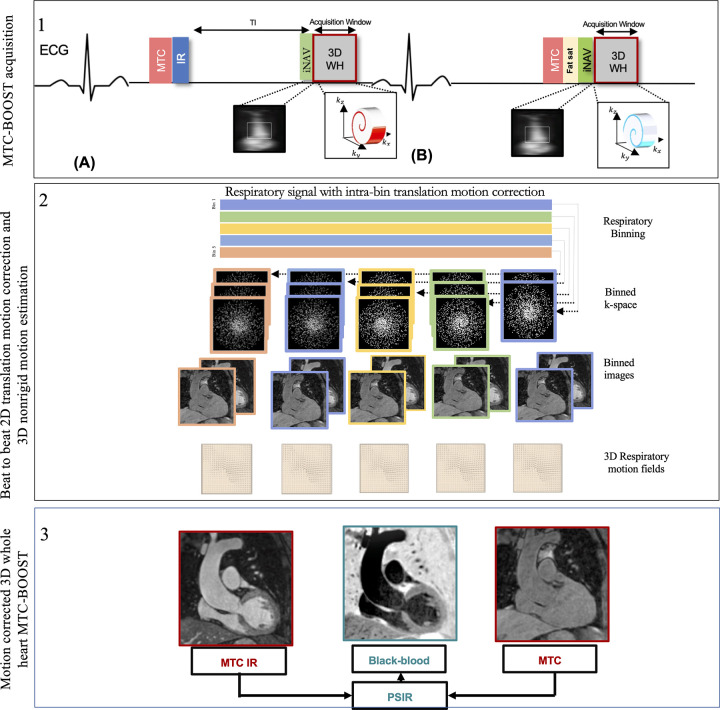
Figure 1:
Schematic overview of investigated free-breathing nonrigid motion-corrected three-dimensional (3D) whole-heart MTC-BOOST framework. Top: Two magnetization-prepared bright‐blood volumes are acquired in odd (A) and even (B) heartbeats. Magnetization transfer in combination with an inversion pulse is used in odd heartbeats, whereas magnetization transfer alone is exploited in even heartbeats. In odd heartbeats, a short inversion time inversion-recovery approach is used to suppress the signal from epicardial fat, whereas frequency‐selective presaturation is used in even heartbeats. Data acquisition is performed using a 3D Cartesian trajectory with spiral profile order. A low‐resolution two-dimensional (2D) iNAV is acquired in each heartbeat by spatially encoding the ramp‐up pulses of the bSSFP sequences. The iNAVs are used to estimate foot-head and right-left rigid motion by tracking a template around the aortic arch, providing motion estimates in a beat-to-beat basis. Middle: Foot-head motion is used to sort the 3D MTC-BOOST data into five equally populated bins, and 3D MR images reconstructed at each respiratory position are used to estimate nonrigid motion between bins. 2D translational beat-to-beat and 3D nonrigid bin-to-bin motion is then integrated into an in-line motion-compensated iterative sensitivity encoding reconstruction to produce the final images. Bottom: The bright‐blood MTC‐IR BOOST and MTC-BOOST volumes are corrected for translation and nonrigid motion and are subsequently combined in a PSIR‐like reconstruction to generate a complementary black‐blood volume. bSSFP = balanced steady-state free precession, ECG = electrocardiography, Fat sat = fat suppression, iNAV = image-based navigator, kx = readout, ky = phase encoding, kz = MRI signal along the scanner bore, MTC-BOOST = Magnetization Transfer Contrast Bright-and-black blOOd phase SensiTive, IR = inversion-recovery pulse, PSIR = phase-sensitive inversion recovery, TI = inversion time, 3D WH = 3D whole-heart.

