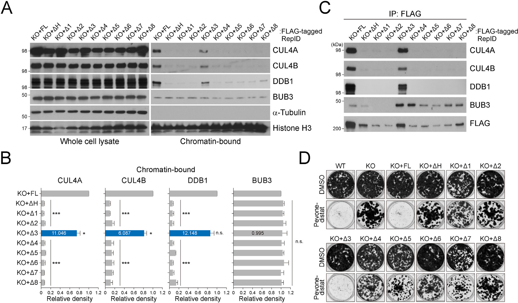
Fig. 2.

CRL4 and its substrate BUB3 occupy different sites of the RepID WD40 domain. (A) CRL4 and BUB3 levels in whole cell lysates and chromatin-bound fractions from U2OS cells transfected as indicated. (B) Quantification of chromatin-bound CUL4A, CUL4B, DDB1, and BUB3 levels. Error bars represent standard deviations from three independent experiments (*p value < 0.05, ***p < 0.001, n.s., not significant, Student’s t-test). Intensity of the exon 3-deletion mutant is indicated as blue bars and inside numbers represent fold change compared to average intensities of CRL4 and BUB3 in other mutants. (C) Immunoprecipitation assay using a FLAG antibody with chromatin-bound fractions was performed and co-precipitated CRL4 and BUB3 were analyzed via immunoblotting. (D) Colony-formation assay with U2OS cells transfected with various RepID mutants in untreated or MLN4924 (pevonedistat)-treated condition.
